
Dog health: oncology
Osteosarcoma in dogs in India: why size beats breed, what amputation really costs a dog, and what three legs actually look like
An Indian owner told their large dog needs a leg removed will assume it is cruel. The evidence says otherwise, and it also says amputation on its own buys limited time. This page gives both with their denominators, names the dogs for whom amputation is the wrong operation, and prices the surgery from a government rate card.
Quick answer
Osteosarcoma in dogs is an aggressive bone cancer, and the first sign is almost always a limp on one leg of a big dog that worsens over weeks. Size drives the risk more than breed: dogs of 50 to 60 kg carried 43 times the odds of a 10 to 20 kg dog. Amputation stops the pain but buys a median 19 weeks alone, against 284 days with chemotherapy. Most dogs cope well on three legs. Your nearby vet decides.
This guide is general information for Indian pet owners and is not a diagnosis. Always consult a registered veterinarian near you before acting on anything you read here, and treat any sudden change in your pet's condition as urgent.
Signs of Osteosarcoma in dogs
The 10 signs to watch for are listed below. Any one of them is a reason to consult your nearby vet; none of them is a diagnosis.
- Limping on one leg that does not settle with rest
- Lameness that gets worse over days to weeks, not better
- A firm, painful swelling over one bone, usually near a joint
- Pain when the leg is touched or the joint is flexed
- Muscle wasting on the affected leg
- Reluctance to jump, climb stairs or walk as far
- Whimpering, restlessness at night, or new irritability
- Reduced appetite and weight loss as the pain drags on
- A sudden fracture after trivial knocks or a small fall
- Swelling or pain at the jaw, ribs, spine or pelvis in axial disease
Breeds on this site that document this condition
Read the denominators before you compare these ten cards, because three different kinds of measurement are sitting side by side. The Great Dane, Rottweiler, Deerhound, Greyhound and Labrador figures are one-year prevalences and odds ratios from a single UK primary-care population of 905,552 dogs, so they describe the chance of a diagnosis being recorded in one calendar year, not a lifetime risk. The Irish Wolfhound, Leonberger and Newfoundland figures are lifetime rates from an owner survey of dogs already old enough to have died, which is why they look so much larger. The Saint Bernard figure comes from a pathology registry, where the denominator is biopsies submitted rather than dogs alive. The Mastiff figure is a share of cancer deaths, not a risk of getting the disease. No Indian study gives a breed breakdown at all, so every card here describes a European population.
The breeds carrying the highest measured odds of osteosarcoma are the large dogs Indian cities actually buy, with the Great Dane at 34.24 times a crossbred's odds, the Rottweiler at 26.67, the Saint Bernard at 6.6 in a UK pathology registry and the Labrador Retriever at 1.57. India has no published canine osteosarcoma case series: a Europe PMC title and abstract search returned zero records on 27 September 2026 while its controls returned 110 and 47, and the affiliation search returned four records of which two were laboratory artefacts. The nearest Indian denominator is a survey of seven practising veterinarians in which osteosarcoma was 3 of 30 bone-related cases, and which records that affected dogs are often misdiagnosed and given calcium and multivitamin supplements. Diagnosis and surgery are cheap at government institutions, with ICAR-IVRI Izatnagar pricing limb amputation at Rs 400 plus tax, a radiograph at Rs 150 and a cytology at Rs 150, and Madras Veterinary College pricing a major surgery at Rs 1,500 while dispensing hospital drugs free of cost. Specialist oncology is another matter: chemotherapy and radiotherapy for dogs exist at ACTREC in Navi Mumbai and at a private practice in Mumbai and Pune, a single infusion session is published at Rs 5,000 to Rs 25,000 or more, and one documented family drove 35 hours from Dehradun to Pune for the operation.
What osteosarcoma is, and how common it actually is
Osteosarcoma in dogs is a malignant tumour of the bone-forming cells, and it does two harmful things at the same time. It destroys the inside of a bone until the leg hurts constantly, and it seeds the lungs long before anyone knows it is there, with micrometastases present in over 90 percent of dogs by the time the tumour is found. It is the commonest primary bone tumour in dogs, making up roughly 80 to 85 percent of all malignant skeletal tumours.
Start with a denominator rather than an adjective. In UK first-opinion practice, a VetCompass study of 905,552 dogs under veterinary care during 2016 confirmed 331 osteosarcoma cases, a one-year period prevalence of 0.037 percent with a 95 percent confidence interval of 0.033 to 0.041. Measured a different way, 764 of 394,061 insured Swedish dogs developed a bone cancer between 1995 and 2002, which is 0.19 percent of the cohort and an incidence rate of 27.2 dogs per 100,000 each year, against roughly 0.89 cases per 100,000 people a year in the comparable Swedish human population. Canine bone cancer is therefore about 27 times commoner than the human disease.
Those two figures do not contradict each other and they are not interchangeable. One counts diagnoses recorded in a single calendar year across every dog on a practice list; the other counts dogs that developed the disease at any point across an eight-year insured cohort. A page that quotes one and calls it the other is telling you something false about your own dog's chances.
Age is the strongest single predictor, and the number moves with the breed. Median age at diagnosis across all breeds in the UK population was 9.64 years, with an interquartile range of 7.97 to 11.41 years and a full range of 1.20 to 18.01 years. A UK national pathology registry of 2,166 osteosarcoma cases found that dogs from high-odds breeds were diagnosed at a median 8.08 years, interquartile range 6.83 to 10.00, against 10.00 years, interquartile range 8.50 to 12.00, for dogs from low-odds breeds, a difference significant at p below 0.0001. In Norwegian Irish Wolfhounds the median age at diagnosis was 5.5 years. In a giant breed this is a middle-aged disease, not an old-dog disease.
Indian clinical experience puts the age distribution in the same place. In a survey of seven practising veterinarians, 80 percent of osteosarcoma cases occurred in dogs over 7 years old and over half in dogs aged 9 years and above.
Why it is so lethal, in numbers
The tumour in the leg is not what kills most dogs. Roughly 80 percent of dogs with osteosarcoma die because of lung metastasis, and one-year survival after diagnosis and treatment is typically under 45 percent. Where metastatic disease is already visible at the time of diagnosis, median survival time is 76 days. That is the reason a limb operation on its own does not solve the problem, and it is the single most important thing to understand before any money is spent.
There is no cure for osteosarcoma in dogs, and the treatment decisions on this page are about how much good time can be bought and at what cost. Everything below is general information. The dog in front of your own veterinarian is the one whose stage, site, age and other illnesses decide what is right, and that decision is theirs and yours together.
Size, not breed, is what carries most of the risk
The breed lists that ranking pages print are largely a proxy for body size, and one study separates the two directly by building a second model in which adult bodyweight and body shape replace breed entirely. What that model shows is a steep, orderly dose-response on weight and no significant effect of being a purebred at all.
In the 905,552-dog VetCompass population, dogs with osteosarcoma had a median adult bodyweight of 33.04 kg, interquartile range 25.50 to 42.93 kg, against 13.95 kg, interquartile range 8.19 to 25.00 kg, for the dogs without it. When breed was removed from the model and replaced by bodyweight and conformation, the gradient across weight bands ran from 0.15 times the odds for dogs under 10 kg to 43.12 times the odds for dogs of 50 to under 60 kg, against a 10 to under 20 kg baseline, every band significant at p below 0.001.
| Adult bodyweight band | Odds ratio | 95 percent confidence interval |
|---|---|---|
| Under 10.0 kg | 0.15 | 0.05 to 0.43 |
| 10.0 to under 20.0 kg | Baseline | Reference category |
| 20.0 to under 30.0 kg | 3.39 | 2.12 to 5.42 |
| 30.0 to under 40.0 kg | 6.68 | 4.22 to 10.56 |
| 40.0 to under 50.0 kg | 14.54 | 8.89 to 23.78 |
| 50.0 to under 60.0 kg | 43.12 | 24.52 to 75.82 |
| 60.0 kg and over | 36.62 | 16.35 to 82.01 |
Two results in that same model deserve more attention than they get. Being a purebred dog carried an odds ratio of 1.15 against crossbreds with a confidence interval of 0.87 to 1.52 and p equal to 0.329, and being a Kennel Club recognised breed carried 1.12, confidence interval 0.87 to 1.45, p equal to 0.378. Neither is significant. Pedigree status is not the risk factor. Mass is.
Body shape adds to it independently of weight. Compared with mesocephalic breeds, dogs from dolichocephalic breeds with long skulls carried 2.72 times the odds, confidence interval 2.06 to 3.58, while brachycephalic breeds carried 0.50, confidence interval 0.32 to 0.80. Chondrodystrophic short-legged breeds carried 0.10 times the odds of non-chondrodystrophic breeds, confidence interval 0.06 to 0.15. Long legs and a long skull raise it; short legs and a short face lower it.
The finding an Indian owner can actually act on is the within-breed one. Dogs at or above the mean bodyweight for their own breed carried 1.65 times the odds of dogs below it, confidence interval 1.27 to 2.13, in the breed-adjusted model, and 2.03 times the odds, confidence interval 1.57 to 2.62, in the other. A heavy Labrador is at higher risk than a lean Labrador. That is the one dial in this disease you turn yourself, and it is the same dial that governs joint disease and heat tolerance in the same dogs.
The same analysis quietly corrects a claim repeated everywhere. Male dogs have long been reported to be at higher risk, and that association disappeared once bodyweight relative to the breed average was taken into account, because male dogs are typically heavier than females of the same breed.
Two giant breeds of the same size, six times the risk
Size explains a great deal and it does not explain everything, and the cleanest demonstration is a Norwegian population survey that followed four breeds born between 1989 and 1998. Among the responders, Irish Wolfhounds recorded 15 primary bone tumours in 169 dogs and Leonbergers 22 in 381, rates of 126 and 72 cases per 10,000 dog-years at risk, while Newfoundlands recorded 4 in 427 and Labrador Retrievers 2 in 938, rates of 11 and 2 per 10,000 dog-years, a difference significant at p below 0.0001.
The Newfoundland and the Leonberger are the same kind of dog by the scales. The authors say so in their own conclusion, that pursuing a search for risk factors other than body size and weight is supported by the significantly different risks of developing primary bone tumours between similarly statured dogs. Genetics is doing the rest of the work. Scottish Deerhounds carry a reported prevalence around 15 percent with a heritability of 0.69, and Deerhounds carrying a putative high-risk dominant allele are considered to have greater than 75 percent risk compared with those not carrying it, while four loci have been associated with osteosarcoma in Irish Wolfhounds and 15 in Rottweilers.
One risk that has nothing to do with size at all
A historical cohort of 683 Rottweilers living in North America, followed for 71,004 dog-months, diagnosed bone sarcoma in 12.6 percent of the dogs. Male and female dogs gonadectomised before 1 year of age had an approximate one in four lifetime risk of bone sarcoma and were significantly more likely to develop it than dogs that stayed sexually intact, with a relative risk of 3.8 and a 95 percent confidence interval of 1.5 to 9.2 in males and 3.1, confidence interval 1.1 to 8.3, in females. The trend test showed a highly significant inverse dose-response between duration of lifetime gonadal exposure and bone sarcoma incidence, at p equal to 0.008 in males and 0.006 in females.
The line that matters most in that paper is the one nobody quotes. The association was independent of adult height or body weight. This is not the size effect wearing a different hat. Read it carefully before you act on it, though, because it is one breed, one questionnaire-based cohort and one outcome, and sterilisation prevents pyometra, mammary disease and unwanted litters that the same dog would otherwise face. The trade-offs, and the Indian timing question, belong on the dog sterilisation in India guide and on the Rottweiler page rather than here. What this page says is narrower. If you own a giant or a high-odds breed, the timing of that routine surgery is a conversation worth having with your vet rather than a date on a calendar.
Where it appears, and why the site changes the conversation
Osteosarcoma is overwhelmingly a disease of the limbs, with up to 80 percent of canine cases in the appendicular skeleton. Within the limbs the distribution is not random, and the pattern is worth memorising because it tells a general practitioner where to point the x-ray machine.
In dogs the forelimbs are affected about twice as often as the hind limbs, and the commonest sites are the distal radius and the proximal humerus, followed by the distal and proximal femur and the distal tibia. Put a hand on your dog and the pattern is easy to hold. The four commonest sites sit away from the elbow and towards the knee, at the growth plates that closed years ago, in the metaphyseal regions of the long bones.
Indian clinical experience gives almost the same ranking with its own numbers. Across the caseload reported by seven practising veterinarians, the sites were radius and ulna at 26.8 percent, femur at 24.8 percent, tibia at 20.3 percent, humerus at 16.1 percent and other bones including the pelvis and mandible at 12 percent.
| Site | Indian veterinary caseload | What the wider literature says |
|---|---|---|
| Radius and ulna, at the wrist end | 26.8 percent | The commonest single site; also the one with the lowest hazard of metastasis |
| Femur | 24.8 percent | Distal femur, at the knee, is a classic hind-limb site |
| Tibia | 20.3 percent | Proximal tibia, at the knee, carries high metastatic ability |
| Humerus | 16.1 percent | Proximal humerus, at the shoulder, is a significant negative prognosticator in meta-analysis |
| Other, including pelvis and mandible | 12 percent | Axial osteosarcoma is less specific to large breeds and is seen in smaller dogs |
Whether the site changes the outlook is genuinely contested, and all three published positions belong on the page. The 162-dog amputation-alone series found no statistically significant relationship between survival time and the site of the primary tumour, whether it was proximal or distal, or whether it was in the forelimb or the hind limb. A meta-analysis of the prognostic-factor literature reached the opposite conclusion, finding a proximal humeral location and an elevated serum alkaline phosphatase both significant negative prognosticators for survival time and for disease-free interval. A treatment review sits between them, reporting that tumours at the distal radius carry a lower hazard of metastasis while those at the proximal humerus, distal femur and proximal tibia metastasise readily, with a resulting increase in mortality. Ask your own vet which position they are working from and why.
Why a pathology registry describes a different disease
A UK national pathology registry recorded 4,847 osteosarcomas among 130,998 histologically confirmed tumours between 2010 and 2023, which is 0.7 percent of all reported tumours. Among the osteosarcomas with a site recorded, long bones accounted for 26.6 percent, the oral cavity for 23.4 percent and the mammary gland for 9.3 percent, and the distribution differed sharply between high-odds and low-odds breeds, with a chi-square of 237.32 on 35 degrees of freedom at p below 0.0001. Long bones dominated in high-odds breeds while oral and mammary tumours were commoner in low-odds breeds.
That looks like a flat contradiction of the 80 percent appendicular figure, and it is not. A pathology registry counts what reaches a laboratory. A classic distal-radius osteosarcoma in a Rottweiler is often diagnosed on radiographs and treated by amputation, with the limb going for confirmation or not at all, while an oral mass is always excised and always submitted. The registry is measuring submission behaviour as much as biology, and the honest reading is that both numbers are right about different questions.
One more site pattern is worth knowing because it is preventable in part. Osteosarcoma has developed at the site of a previously healed fracture, in chronic osteomyelitis, in association with metallic implants and after exposure to radiation therapy given for something else. It has also been reported concurrently with bone infarcts. A dog with an old orthopaedic plate that becomes lame at that exact spot years later deserves a radiograph, not reassurance.
What you will notice, and the week a limp stops being a limp
The first sign is a limp on one leg in a big dog that does not settle with rest, and that gets worse over days to weeks rather than better. Alongside it you may find a firm, localised swelling over the bone and loss of muscle on that leg. That combination is a this-week radiograph, not a wait-and-see.
The reason cases are caught late is that the early picture is ordinary. Early signs are subtle and easily mistaken for minor injuries, and unlike a soft-tissue injury the pain progressively worsens over time rather than fading. Indian clinicians describe the same trap, with signs often misattributed to orthopaedic disorders such as arthritis or trauma in the early stages, so that by the time a definitive diagnosis is established many dogs already harbour micrometastatic disease in the lungs even when nothing is visible on radiographs.
One Indian case shows the cost of that delay in calendar time. An Indian cross-breed dog named Badal began limping and panting at the end of 2022 and was given injections that did not improve his condition; by the end of August the following year the limping was noticeable and he had a fall; a local vet raised the possibility of cancer after an x-ray; a biopsy, a CT scan and a fine-needle aspirate in October confirmed a tumour; and the amputation was performed on 9 January 2024. That is roughly a year from the first limp to the operation.
- A limp that has not improved after a week of rest in a dog over 20 kg earns a radiograph, because the weight bands above 20 kg are where the odds ratios climb steeply and because the pain of bone cancer worsens rather than settles.
- A firm swelling you can feel over a bone beats a swelling over a joint, since the tumour destroys normal bone and replaces it with cancerous bone that bulges.
- Do not accept a second course of painkillers without imaging. Lameness in a young adult of a high-odds breed should be treated as an osteosarcoma question until radiographs say otherwise, because high-odds breeds are diagnosed at a median 8.08 years against 10.00 in low-odds breeds.
- A leg that breaks after a small knock is not clumsiness. As the tumour grows it weakens the bone until it is susceptible to breaking, and a pathological fracture in a large dog is a same-day problem.
- Reduced appetite, weight loss and night-time restlessness are pain, not ageing, and they were among the advanced signs recorded in Indian clinical practice alongside lethargy and withdrawal.
Do not let a big dog's one-legged limp be treated blind for a second time. The commonest way an Indian dog loses its chance at a good outcome is a second or third course of anti-inflammatory injections given without a radiograph of the painful bone, because early osteosarcoma looks exactly like a strain and the pain settles a little each time. Ask for a radiograph of the specific bone that hurts, and ask for chest radiographs at the same visit. If your dog cannot bear weight at all, is crying, or the leg has changed shape suddenly, treat that as an emergency and find a registered veterinarian today rather than waiting for an appointment. What happens next is your nearby vet's decision for your dog, not this page's.
Two things commonly mistaken for osteosarcoma are worth naming too, because the mistake runs in both directions. Elbow dysplasia produces a chronic forelimb lameness in exactly the breeds this page is about, and it is covered on the elbow dysplasia in dogs page. And a benign expansile bone lesion can look malignant on imaging, which is the subject of the next section.
How it is confirmed, and why the biopsy is not a formality
A radiograph of the painful bone is the first test, and it is often enough to make an experienced veterinarian confident. Confidence is not a diagnosis. A biopsy of the tumour is the only way to diagnose osteosarcoma definitively, and on an Indian budget it is also the cheapest part of the whole exercise.
The radiographic hallmarks are aggressive osteolysis, cortical thinning, periosteal proliferation with a characteristic sunburst pattern and irregular new bone formation. A definitive diagnosis requires histopathological confirmation, usually through a core biopsy; a fine-needle aspirate can give preliminary cytological information but lacks sufficient accuracy compared with histopathology. Alongside that, chest radiographs, blood work and a urinalysis are taken to see how far the disease has already travelled and whether the dog is well enough for what comes next.
Most dogs are already at stage IIB when they are diagnosed, meaning a high-grade tumour that has extended beyond the bone into nearby structures without detectable metastasis. That is not a failure of owner vigilance. It is what a disease that spreads microscopically before it is visible looks like from the outside.
The Indian case that shows why the biopsy earns its money
In 2026 the Animal Oncology Group at ACTREC, Tata Memorial Centre in Navi Mumbai published the case of a 10-year-old neutered female German Shepherd Dog weighing 31 kg, presented with a three to four week history of progressive right forelimb lameness and swelling localised to the distal radius, with no known history of trauma. Clinical examination found intermittently non-weight-bearing lameness and a firm, painful swelling over the radial metaphysis, and computed tomography showed an expansile lytic lesion affecting the distal radius and involving the articular surface with peripheral sclerosis and cortical erosion. The differential diagnosis included osteosarcoma, giant cell tumour and aneurysmal bone cyst.
That is the commonest osteosarcoma site in the commonest osteosarcoma presentation in a large breed. The radiographs argued against cancer, showing sharp lesion margins, a smooth zone of transition and no periosteal reaction, and thoracic imaging showed no metastatic spread. A bone biopsy taken from the wall of the lesion found fibrous tissue and osteolytic change with no giant cells and no neoplastic cells, confirming an aneurysmal bone cyst. The dog was treated with percutaneous sclerotherapy under general anaesthesia, which the authors note avoided the morbidity associated with curettage, bone grafting or limb amputation.
The outcome is the part to carry away. Radiographs at eight weeks showed healing of the affected area, and at the seven-month examination the dog had regained normal symmetrical forelimb muscle tone with no lameness at walk or trot and a pain score of zero; the owner later reported that the dog lived 13 months after treatment and died of renal insufficiency and haemorrhagic enteritis with no lameness up to the end. The authors' own framing is the lesson. Aneurysmal bone cysts are rare in dogs and often mimic malignant bone tumours on imaging, and differentiating them from osteosarcoma is essential yet challenging because both can present with cortical destruction, lytic changes and giant-cell-rich cytology. One dog, one biopsy, one leg kept.
What the report will and will not tell you
Two pieces of the report change what you should expect and one does not. An elevated serum alkaline phosphatase is a significant negative prognosticator for both survival time and disease-free interval in meta-analysis, and it is a line on an ordinary biochemistry panel. Tumour location, specifically the proximal humerus, is the other significant negative prognosticator in that same analysis.
The histological subtype, by contrast, appears not to matter for outcome. In 57 dogs with surgically resected appendicular osteosarcoma between 2014 and 2022, 43 had osteoblastic tumours and microvascular invasion was evident in 5; there was no significant difference in progression-free interval or overall survival between subtypes, and dogs with microvascular invasion had a median overall survival of 168 days against 262 days for those without, which was not significant. If a pathology report names a subtype you have never heard of, that is a description rather than a prognosis.
- Ask for a radiograph of the exact bone that hurts, not a general limb film, because the lesion is metaphyseal and a wide view can bury it.
- Ask for chest radiographs at the same visit. They change the whole conversation, since visible metastasis at diagnosis carries a median survival of 76 days.
- Ask whether a biopsy is possible before the limb is removed. A biopsy of the tumour is the only definitive diagnosis, and a published Indian case at this exact site turned out to be a benign cyst.
- Ask for the serum alkaline phosphatase result specifically, because an elevated value is one of only two prognostic factors that survived meta-analysis.
- Ask what the plan is if the biopsy is inconclusive. Cytology gives preliminary information and histopathology is the standard, so a first sample that does not answer the question is a normal event rather than a wasted one.
Amputation, honestly: what dogs on three legs actually do
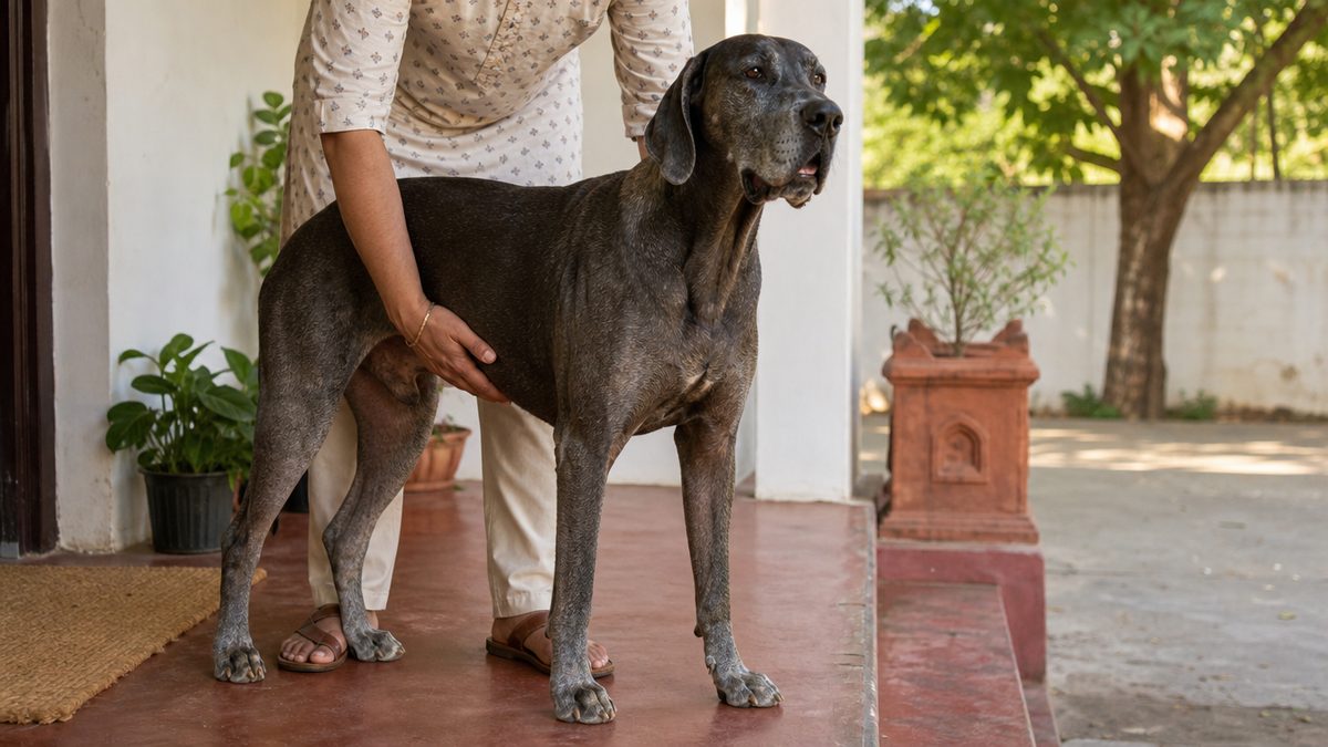
Removing the leg is the standard local treatment for appendicular osteosarcoma, and it is done to take out the primary tumour, to stop the pain and to remove the risk of the bone breaking. For a forelimb lesion the whole forequarter is generally removed, and for a hind limb the amputation is made at the hip. An Indian owner hearing that will usually assume it is cruel. The published outcome studies say something different, and they also say clearly what amputation on its own does not do.
The largest owner-outcome series followed 64 dogs that had a thoracic or pelvic limb amputated at a veterinary teaching hospital between 2005 and 2012, with owners interviewed by telephone afterwards. Fifty-eight of the 64 owners, which is 91 percent, perceived no change in their dog's attitude after amputation; 56, or 88 percent, reported a complete or nearly complete return to the dog's quality of life before surgery; 50, or 78 percent, said recovery and adaptation were better than they had expected; and 47, or 73 percent, reported no change in the dog's recreational activities. Taking everything into account, 55 of 64 respondents, 86 percent, said they would make the same decision again, 4 said they would not, and 5 were unsure.
An older Dutch study interviewed 44 owners and found that most dogs adapted to walking on three legs within a month, sooner than most of the owners had expected, with few complications after surgery, although changes in behaviour were observed in approximately one-third of the dogs. That last clause matters and is usually left out. A third of owners noticed their dog was different in some way afterwards, and you should be told that before you consent rather than after.
What the gait laboratory measured, rather than what owners felt
Twelve dogs weighing between 8 and 40 kg had force-plate and kinematic gait analysis before a hind limb amputation and again at 10, 30, 90 and 120 days afterwards, with magnetic resonance imaging of the opposite stifle before and at 120 days. The load redistributed across all the remaining limbs, and by ten days after the procedure the dogs had already established their new locomotory pattern. Ranges of motion were very similar before and after the amputation and stayed constant across the later sessions.
The most useful result answers the fear owners actually have, which is that the remaining legs will be wrecked. There were no changes in the signal intensity of the soft tissues evaluated and no evidence of cartilage damage or osteoarthritis on magnetic resonance imaging of the opposite stifle at 120 days. The authors also found that the adaptation process began before the amputation was performed, because a dog with a painful leg has already stopped using it.
Indian practice reports the same speed. Badal's amputation in Pune lasted two and a half hours without complications and the dog was able to walk within 24 hours of surgery.
The giant-breed weight question, which is not what the folklore says
The common Indian objection is that a Great Dane or a Saint Bernard is simply too heavy to manage on three legs. That is not what the outcome studies found, and it is not nothing either. The 64-dog series recorded that body condition score and body weight at the time of amputation were negatively correlated with quality of life scores after surgery, and the authors' stated clinical conclusion was the need for postoperative weight control in these patients. The gait study included a 39 kg Swiss Mountain Dog and a 40 kg Bernese Mountain Dog among its twelve, and reported quick adaptation across the group. In the 165-dog palliative radiotherapy cohort, weight was among the factors that were not associated with survival time.
Read together, those three say that weight is a quality-of-life variable to manage rather than a disqualification to accept. A heavy dog can be an amputee. A heavy dog that is also overweight will do worse than the same dog at a lean body condition, and the weeks before surgery are the time to start on that, not the weeks after.
When amputation is the wrong operation
There is a defined group of dogs for whom limb-sparing surgery is preferred over amputation, and it is not a matter of owner preference. Most dogs perform well with an amputation, but where the dog suffers from severe pre-existing neurological and orthopaedic disease, limb-sparing is preferred. A treatment review says the same thing from the other direction, recommending limb-sparing for dogs with compromising neurologic or orthopedic problems, or where owners refuse amputation. A dog that already cannot use another leg properly cannot spare the one you are proposing to remove.
Limb-sparing is not a soft option and the complication rates are why. Limb function is preserved in over 80 percent of dogs after limb-sparing surgery, but infection occurs in 30 to 50 percent of patients, implant failure in 20 to 40 percent and tumour recurrence in 15 to 25 percent of cases. The candidate criteria are narrow, requiring a primary tumour confined to the bone, otherwise good general health, no pathological fracture, less than 360-degree involvement of the surrounding soft tissues and a firm rather than an oedematous soft-tissue mass. Survival is not the trade-off being made, because canine reports suggest a similar metastatic frequency and survival time with limb salvage compared with amputation and adjuvant chemotherapy.
In India the practical position is narrower still. Limb-sparing needs an endoprosthesis or a cortical allograft or a circular external fixator, a surgeon who does the procedure regularly and the ability to manage an infection rate of up to half. Where that is not obtainable, the honest choice is between amputation and pain control, and the next two sections are about both.
What amputation alone buys, and what chemotherapy adds
Amputation on its own is a pain operation, not a cancer operation. It removes the primary tumour and the fracture risk but does not prevent the spread of the cancer, because the lungs are already seeded. The number that proves it is 34 years old and every survival figure you will read traces back to it.
Long-term follow-up on 162 dogs with appendicular osteosarcoma treated by amputation alone, collected from 17 veterinary institutions, gave a mean survival of 19.8 weeks and a median of 19.2 weeks, with one-year and two-year survival rates estimated at 11.5 percent and 2.0 percent. The majority, 72.5 percent, died or were euthanased because of problems documented to be related to metastases, with the first clinically apparent sites being the lungs in 60.8 percent of the total, the skeleton in 5.2 percent and both in 4.6 percent. Cornell states the same thing in one line for owners, that the median survival time with amputation alone is four months.
Adding chemotherapy roughly doubles to trebles that, and the largest single-population comparison puts real confidence intervals around it. Among 470 dogs treated with amputation followed by one of five carboplatin or doxorubicin-based protocols between 1997 and 2012, the overall median disease-free interval was 291 days with a 95 percent confidence interval of 253 to 362 days, and the overall median survival time was 284 days, confidence interval 248 to 316 days. For the dogs that actually completed the protocol, the median disease-free interval was 398 days, confidence interval 317 to 492, and the median survival 367 days, confidence interval 325 to 401.
That gap between 284 and 367 days is the most practical number on this page for an Indian family. It is the difference between starting a course and finishing it, and it is roughly three months of a dog's life. Before you begin, ask what the whole course costs and whether you can see it through.
| Approach | What was measured | Population |
|---|---|---|
| No treatment | Pain relief only; the disease itself is untouched | Not separately measured in the studies opened for this page |
| Amputation alone | Median survival 19.2 weeks, one-year survival 11.5 percent, two-year survival 2.0 percent | 162 dogs across 17 institutions, 1978 to 1988 |
| Amputation plus chemotherapy, as prescribed | Median survival 284 days, median disease-free interval 291 days | 470 dogs, 1997 to 2012, intention to treat |
| Amputation plus chemotherapy, protocol completed | Median survival 367 days, median disease-free interval 398 days | The subset of those 470 dogs that finished the course |
| Amputation plus single-agent carboplatin | Median survival 307 days against approximately 138 days for surgery alone | 48 dogs, as reported in a treatment review |
| Amputation plus doxorubicin | Median survival 366 days against 138 days in a historical control group | 35 dogs against 162 historical controls |
| Palliative radiation, no amputation | Median survival 119 days with bisphosphonates and 109 days without, p equal to 0.758 | 165 dogs, 2010 to 2019 |
Read that table as a ranking of treatments and you will misread it. These are medians from different decades, different countries and different case mixes rather than a head-to-head trial, and the protocol choice itself appears to matter less than finishing. After adjustment for baseline characteristics and prognostic factors, none of the five protocols in the 470-dog cohort gave a significant reduction in the risk of metastasis or death, and dose intensity was not associated with either outcome. What did differ was tolerability, with a lower proportion of dogs on the six-dose carboplatin protocol experiencing adverse events, 48.4 percent against 60.8 to 75.8 percent on the others, which the authors note could be an advantage in maintaining quality of life during treatment.
Two-year survival is where the popular pages and the measured data part company. The Indian page that ranks first on the bare query states that approximately 20 to 25 percent of dogs survive beyond two years with amputation plus chemotherapy. In the 470-dog cohort the two-year survival proportions by protocol ran from 0.13 to 0.20, and the three-year proportions from 0.03 to 0.10. Both are printed here with their populations named. The measured figure sits at the bottom of the quoted range or below it.
Immunotherapy and the newer agents are not an option for a dog in India today. Listeria-based vaccines, dendritic cell therapy and tyrosine kinase inhibitors such as toceranib show promise and remain largely confined to research or referral centres. No Indian source opened for this page names any of them as obtainable here, so treat them as unavailable until a supplier says otherwise.
Pain control is the floor of care, not the fallback
A dog whose owner declines surgery still has a painful bone tumour, and that dog still deserves a written analgesia plan. Where surgery is not an option, pain management becomes the focus of care rather than the absence of it. The AAHA oncology guidelines put pain management alongside chemotherapy, immunotherapy, radiation therapy, surgery and nutritional support as part of the same multimodal therapy, not as what is left over.
The drug classes used are non-steroidal anti-inflammatory drugs, gabapentin, amantadine and opioids, alone or in combination, and the choice among them is your veterinarian's. Indian clinical practice describes the same set plus bisphosphonates such as pamidronate and zoledronate, which inhibit osteoclastic bone resorption. This page names classes and never an amount, because the amount is a decision made for one animal by the person examining it.
The bisphosphonate deserves a harder look than the ranking pages give it, because the evidence has moved and they have not. A prospective study of 43 dogs given single-agent pamidronate with a non-steroidal anti-inflammatory found that 12 of the 43, 28 percent, had pain alleviation for more than four months, lasting a median of 231 days, with statistically significant differences in pain index and bone mineral density between responders and non-responders. That is a real subset and a real benefit.
Then the same group ran a randomised, prospective, double-blinded, placebo-controlled trial in 50 dogs, 26 given adjuvant pamidronate and 24 given sterile saline on top of standardised palliative therapy. Median duration of subjective pain relief was 76 days with pamidronate and 75 days with placebo, at p equal to 0.39. Forty percent of the dogs, 20 of 50, achieved durable analgesia lasting 112 days or more, made up of 11 of 26 on pamidronate and 9 of 24 on placebo. The authors concluded that combining pamidronate with standardised palliative therapy is safe but does not clearly improve pain alleviation, while in the dogs that did achieve durable pain control it did reduce focal bone resorption.
The larger retrospective agrees. Among 165 dogs given hypofractionated palliative radiation therapy between 2010 and 2019, of whom 68 also received bisphosphonates, median survival from the first radiation treatment was 119 days in the bisphosphonate group and 109 days without, a difference that was not significant at p equal to 0.758. Only age over 9 years was prognostic in that population, at p equal to 0.031, while bisphosphonate drug type, timing, tumour location, weight, breed, sex, time to treatment, concurrent chemotherapy and salvage amputation were all unassociated with survival.
Declining amputation is a treatment decision, not the absence of one. Ask your vet for a written comfort plan naming the drug classes, the review interval and the specific changes that would mean the plan is failing, and ask what to do at two in the morning when it does. Weigh the dog at every visit and write it down. Restrict slippery floors, stairs and jumping, because the weakened bone is susceptible to breaking with minimal trauma. Agree in advance what you will do if the leg fractures, since that is the single event most likely to force a decision at speed. Palliative radiation and intravenous bisphosphonate therapy are further options where a specialist is reachable. Your nearby vet decides what is right for your dog, and a plan made calmly at diagnosis is kinder than one made in a crisis.
Palliative radiation is the option most Indian owners will not be offered and should still know about. It is considered a palliative method whose intent is to provide pain relief and prolong life, it is not easily accessible and it requires a general anaesthetic for each session. In a 50-dog study, median survival was longest for dogs receiving radiotherapy together with chemotherapy at 307 days and shortest for those receiving radiotherapy with pamidronate at 69 days. Adding metronomic lomustine to a two-fraction palliative radiation protocol in 86 dogs gave a median survival of 184 days against 154 days without it, which was not significant at p equal to 0.84. The Indian page that ranks first describes palliative radiation as providing significant pain relief for two to four months in many cases.
What it costs in India, and what is actually obtainable
The surprising part of the Indian cost picture is that the operation is cheap and the drugs afterwards are not. Two government veterinary institutions publish their rate cards, and one of them prices limb amputation by name. No ranking page opened for this article prints a rupee figure for diagnosing or treating this disease.
The ICAR-Indian Veterinary Research Institute at Izatnagar publishes a surgical rate card effective June 2024 for its Referral Veterinary Polyclinic, with a column for pet animals and a footnote reading plus tax as applicable. Line 38 of that card is Limb Amputation, priced at Rs 400 for a pet animal. On the same card, plain radiography is Rs 150, ultrasonography Rs 250 and chemical restraint Rs 100, while the laboratory card prices fine-needle aspiration cytology at Rs 150, histopathology at Rs 150 and haematology at Rs 300. Registration for a dog or cat is Rs 30 at the normal rate, rising to Rs 200 after 12:30 in the afternoon and Rs 400 after 5 in the evening or on holidays.
At Madras Veterinary College in Chennai, Tamil Nadu, the published Teaching Hospital fee structure is built differently, by procedure class rather than by named operation. A small animal registration valid for seven days is Rs 100, a major surgery is Rs 1,500, a specialised surgery Rs 3,000 and an emergency surgery Rs 2,500, with an x-ray at Rs 100, a scan at Rs 300, a plain computed tomography scan with report at Rs 2,500 and haematology, biochemistry, urine or culture at Rs 50. The hospital's pharmacy runs on an average annual budget of Rs 25,00,000 and dispenses drugs and fluids free of cost for the treatment of animals brought to the hospital. Its referral units include Orthopaedics and Radiology and Computed Tomography, and oncology is listed among the faculty disciplines.
Every rupee figure on this page has been divided by 84.38 and by 74 to check it is a native Indian price rather than a converted dollar list. Rs 400 is about 4.74 or 5.41 US dollars on those two rates, Rs 1,500 is about 17.78 or 20.27, and Rs 3,000 is about 35.55 or 40.54. None of them is a round dollar number, so the cards are priced in rupees rather than translated.
| Item | Published price | Source and date |
|---|---|---|
| Limb amputation, pet animal | Rs 400, plus tax | ICAR-IVRI Izatnagar surgical rate card effective June 2024 |
| Excision of tumour, pet animal | Rs 300, plus tax | ICAR-IVRI Izatnagar surgical rate card effective June 2024 |
| Plain radiography, pet animal | Rs 150, plus tax | ICAR-IVRI Izatnagar surgical and imaging rate card |
| Fine-needle aspiration cytology, pet animal | Rs 150, plus tax | ICAR-IVRI Izatnagar laboratory rate card |
| Histopathology, pet animal | Rs 150, plus tax | ICAR-IVRI Izatnagar laboratory rate card |
| Chemical restraint, pet animal | Rs 100, plus tax | ICAR-IVRI Izatnagar surgical rate card |
| Major surgery | Rs 1,500 | Madras Veterinary College Teaching Hospital fee structure |
| Specialised surgery | Rs 3,000 | Madras Veterinary College Teaching Hospital fee structure |
| Plain computed tomography with report | Rs 2,500 | Madras Veterinary College Teaching Hospital fee structure |
| Haematology, biochemistry, urine or culture | Rs 50 | Madras Veterinary College Teaching Hospital fee structure |
| One chemotherapy infusion session at a specialist centre | Rs 5,000 to Rs 25,000 or more, depending on drug and facility | VOSD cost table headed Price Range (INR), 15 June 2026 |
| Canine cancer care, chemotherapy or radiation, ACTREC Kharghar | Rs 40,000 to Rs 60,000 depending on cycles | ACTREC press item, 24 June 2015 |
Set the surgery against the drugs and the shape of the problem is obvious. A government institute prices the amputation itself at Rs 400 plus tax, while a single chemotherapy infusion session at an Indian specialist oncology centre is published at Rs 5,000 to Rs 25,000 or more by an animal charity in Bengaluru, Karnataka. Cornell describes the standard adjuvant course as an intravenous drug given every three weeks for four to five months, so the session figure is the one that multiplies. This page does not print a total, because no Indian source publishes a total for an osteosarcoma chemotherapy course and a number this page calculated would not be a quotation from anybody.
Two caveats belong with the rate card before anyone books a train ticket. A line on a government surgical card covers the procedure, not the anaesthesia, the hospitalisation, the imaging or the drugs, all of which are separate lines or separate cards, and those cards are written for animals presented at those institutions rather than as a national tariff. A private Indian practice will quote its own figure and is entitled to. What the cards prove is narrower and still useful, which is that the amputation is not the expensive part, so cost is the wrong reason to refuse it.
Pet insurance wordings differ sharply on whether an oncology claim of this shape is covered at all, and they are compared on the pet insurance in India guide rather than here.
What is different about osteosarcoma in India
Three things change in India, and none of them is biology. The dogs that carry the highest risk are exactly the dogs Indian cities buy, the specialist care that follows an amputation exists at a handful of addresses, and there is almost no published Indian evidence to argue from.
Start with the breeds. The highest-odds breeds in the UK population are the Scottish Deerhound, Leonberger, Great Dane and Rottweiler, and of those the Great Dane and the Rottweiler are ordinary sights in Indian apartments. Add the Saint Bernard at 6.6 times the odds of a crossbred in a UK pathology registry and the Labrador Retriever, India's most popular large dog, at 1.57 times the odds and 11.48 percent of an entire national caseload, and the Indian exposure is real rather than theoretical. Indian veterinarians name the same set, listing Rottweilers, Great Danes, Saint Bernards, Irish Wolfhounds and Scottish Deerhounds as showing particularly high incidence.
The Indian evidence, stated precisely
There is no published Indian case series of canine osteosarcoma that this page could find, and the search behind that sentence is worth printing rather than asserting. A Europe PMC title-and-abstract search for osteosarcoma combined with dog, dogs or canine and India returned 0 records on 27 September 2026, and the author-affiliation search returned 4. Two of those four are laboratory artefacts, one a protein-interaction study in human and canine osteosarcoma cell lines with an Indian co-author, and one a polymer-chemistry paper on iron sensors that matched because it imaged a human osteosarcoma cell line and a canine kidney cell line. Controls run through the identical route in the same session returned 110 records for Labrador with hip dysplasia and 47 for parvovirus in dogs in India, so the instrument was working and the absence is real.
The two records that are not artefacts are both worth having. One is the ACTREC aneurysmal bone cyst case described further up this page, an Indian dog with a distal radial lesion whose differential included osteosarcoma and whose leg was saved by biopsy. The other is an evaluation of a bone-seeking radiopharmaceutical in four canine patients with primary and metastatic skeletal lesions, published by an author list combining Indian and Hungarian groups, in which two of the three dogs with primary bone cancer showed stable disease initially with a palliative effect, one dog with skeletal metastasis from a primary elsewhere responded well, and a fourth with metal-implant-induced osteosarcoma and severe limb oedema did not respond at all.
The nearest thing to an Indian denominator comes from outside the indexed literature. A 2025 mixed-methods study from Ahmedabad surveyed seven practising veterinarians by phone and in person, and reported that each veterinarian saw an average of seven to eight new osteosarcoma cases a year, and that among 30 bone-related clinical cases reviewed across the participants osteosarcoma accounted for approximately 10 percent, or 3 of 30, behind fractures, osteomyelitis and inflammatory bone disease. Thirty cases and seven clinicians is a small, qualitative denominator and it is the only one India has.
That same study carries the most uncomfortable Indian finding on this page. Specialised veterinary facilities capable of diagnosing and treating these cancers are limited, especially in under-resourced regions, and many affected dogs are misdiagnosed or inadequately treated with symptomatic supplements such as calcium and multivitamins, which offer no therapeutic benefit for malignancies. If your large dog is limping and the prescription is a calcium supplement, that is the moment to ask for a radiograph.
Where specialist care actually exists, and what a referral costs in hours
Veterinary oncology in India is a short list of addresses. The Tata Memorial Centre's ACTREC campus at Kharghar in Navi Mumbai was described in its own press item of 24 June 2015 as offering chemotherapy and radiotherapy for canines and as the only such unit in the country, naming Dr Pradip Chaudhari as the veterinary oncologist, recording nearly 150 dogs treated in the preceding year of which nearly 50 underwent chemotherapy, and quoting the observation that most city veterinary doctors are unaware the treatment is available in India at all. That unit is still running: the 2026 bone-cyst case above was published from ACTREC's Comparative Oncology Program and Translational Preclinical Imaging and Radiotherapy Facility, within its Animal Oncology Group.
Every veterinary oncology address named by any source opened for this page sits in one state. ACTREC is at Kharghar in Navi Mumbai, Maharashtra, and the private specialist practice runs in Mumbai and Pune, also Maharashtra The Cancer Vet, Mumbai and Pune. Clinic…. An owner in Delhi, Kolkata, Bengaluru or Hyderabad whose dog needs chemotherapy after an amputation is therefore looking at a journey rather than an appointment, which is the single most important practical fact on this page for anyone outside Maharashtra.
A second address has appeared since. The Cancer Vet runs clinics in Mumbai and Pune offering cancer diagnosis and treatment, chemotherapy, radiation therapy, surgical treatment and tele-consultations, founded by a board-certified veterinary radiation oncologist, and publishes no prices on its site. So the answer to whether palliative radiotherapy for a dog exists anywhere in India is yes, at two named addresses in one state, and the answer to what it costs is that nobody publishes a current figure; the only Indian number in print bundles radiation into a Rs 40,000 to Rs 60,000 range for canine cancer care generally and is eleven years old.
What that geography costs a family was documented in January 2024. An owner in Dehradun drove her twelve-and-a-half-year-old Indian cross-breed dog to Pune for the amputation, a journey of four days and 35 hours of driving with regular breaks, after a local vet's x-ray raised the possibility of cancer. The surgery itself took two and a half hours without complications and the dog walked within 24 hours. Two and a half hours of surgery, four days of travel. That ratio is the Indian problem in one line, and it is why the question to ask your own vet first is whether they can do the amputation locally rather than whether a specialist exists somewhere.
A note on that case worth keeping straight. It is a newspaper report of one dog, with no follow-up survival and no denominator, so it tells you the operation is obtainable in India and nothing about how long the dog lived. Treat it as a demonstration, not as evidence.
The monsoon and the heat matter less to the tumour than to the calendar. An adjuvant course is an intravenous drug given every three weeks for four to five months, which for a family more than a day's travel from one of two cities means four or five long journeys, some of them in a humid monsoon month when the roads are worst and a large dog travels badly. That is a real and legitimate reason to choose amputation with pain control over amputation with chemotherapy, and it is a reason to say so out loud at the first consultation rather than to start a course and abandon it, given that finishing the protocol was worth a median 367 days against 284.
Ask the questions in this order. Ask first whether your own vet can refer you anywhere at all, and to whom by name, because in most Indian cities the honest answer is that there is nobody local to send you to. Ask second what each separate step costs, because the amputation is affordable on a government rate card at Rs 400 plus tax while one infusion session at a specialist centre is published at Rs 5,000 to Rs 25,000 or more Voice of Stray Dogs (VOSD). Ask third what happens if you stop partway, because a course abandoned at week six is not the course any median on this page was measured on.
Living with it, week by week, and deciding when
A dog recovering from an amputation has two jobs, which are to get comfortable and to stay lean, and an owner has a third, which is to watch the lungs. Even with multimodality therapy, metastasis to the lungs often occurs, and rechecks every two to three months to monitor radiographs for metastasis may be recommended.
The recovery timetable from the gait laboratory is faster than most owners expect. Load redistributes across the remaining limbs immediately, a new locomotory pattern is established by ten days after surgery, and ranges of motion are very similar before and after the amputation. Most dogs in the Dutch survey adapted to walking on three legs within a month, sooner than their owners had expected.
Two honest qualifications go with that. Behaviour changes were observed in approximately one-third of the dogs in that survey, and body condition score and body weight at the time of amputation were negatively correlated with quality of life scores afterwards. Keeping a three-legged giant breed lean is not cosmetic. It is the intervention with the clearest published link to how the dog feels a year later.
- Weigh the dog at every visit and write the number down. Body weight at amputation predicted worse quality-of-life scores afterwards, and postoperative weight control was the authors' stated clinical conclusion.
- Book chest radiographs on a calendar, not on a worry. Rechecks every two to three months to monitor for metastasis are the recommended pattern, because the lungs are where this disease goes.
- Take the rugs up and block the stairs for the first month, since dogs establish their new pattern within about ten days and a slip in week one is the commonest avoidable setback.
- Tell your vet the moment a cough, faster breathing or exercise intolerance appears. Roughly 80 percent of dogs with this disease die because of lung metastasis.
- Ask for the pain plan in writing even after the leg is gone, because pain management sits inside the guideline alongside surgery and chemotherapy rather than after them.
Know what the second year looks like before you plan for it. Of the dogs that survive beyond one year, approximately 54 percent will develop metastatic disease, with a median survival time of 243 days from that point. Two-year survival proportions in the 470-dog chemotherapy cohort ran from 0.13 to 0.20 depending on protocol, and three-year proportions from 0.03 to 0.10. A dog that reaches its second year with this disease has done well by the published numbers, and it has not been cured.
The target is comfortable months, not years, and that target is reachable. In the 64-dog owner series, 91 percent saw no change in their dog's attitude after amputation, 88 percent reported a complete or nearly complete return to the quality of life the dog had before surgery, and 86 percent said they would make the same decision again. The opposite stifle showed no cartilage damage or osteoarthritis on imaging at 120 days. Judge the plan on whether your dog eats, sleeps, greets you and moves around the house without pain, and review that judgement with your vet at every visit rather than once at the end. The decision about when enough is enough belongs to you and your nearby vet together, and making it early and in writing is kinder than making it at speed.
Owners searching for the final stages of bone cancer in dogs are usually asking one practical question, which is how they will know. This disease ends in one of two ways. Either the lungs fill, which an owner sees as a cough, faster breathing and a dog that will not walk as far, since roughly 80 percent of dogs with osteosarcoma die because of lung metastasis. Or, in a dog whose leg was not removed, the weakened bone breaks after minimal trauma. Knowing which of those two you are watching for is what lets a family decide in advance instead of at two in the morning.
One last framing that helps families decide. Osteosarcoma is not the only cancer that presents as a lump or a limp in a middle-aged Indian dog, and the numbers are different for each. Lymphoma in dogs and cats presents as painless swollen nodes and responds far better to drugs, and mammary tumours in dogs and cats are the commonest tumour of entire female dogs. Getting the name right before spending anything is the whole point of the biopsy. Older dogs generally, including the ones on three legs, are covered on the senior dog care in India guide.
Common questions about osteosarcoma in dogs
How long does my dog have to live with osteosarcoma?
It depends almost entirely on what is done next. With amputation alone, median survival in 162 dogs was 19.2 weeks, with 11.5 percent alive at one year and 2.0 percent at two. With amputation plus chemotherapy in 470 dogs, median survival was 284 days as prescribed and 367 days for the dogs that completed the course.
What are the first signs of osteosarcoma in dogs?
A limp on one leg of a large dog that does not settle with rest and worsens over days to weeks, usually with a firm painful swelling over one bone near a joint and muscle wasting on that leg. Early signs are subtle and easily mistaken for a strain, which is why many cases are found late.
What is the deadliest cancer in dogs?
Osteosarcoma is among the most lethal. It is the commonest primary bone tumour, roughly 80 to 85 percent of malignant skeletal tumours, about 80 percent of affected dogs die of lung metastasis, and micrometastases are already present in over 90 percent at diagnosis. One-year survival after diagnosis and treatment is typically under 45 percent.
What are 5 symptoms of osteosarcoma?
Five are worth learning. One, limping on one leg that worsens rather than settles. Two, a firm painful swelling over a bone. Three, pain when that spot is touched. Four, muscle wasting on the affected leg. Five, a sudden fracture after a trivial knock, because the tumour weakens the bone from inside.
What is the life expectancy of a dog with bone cancer in the leg?
Published medians run from about 19 weeks with amputation alone to 284 days with amputation plus chemotherapy in 470 dogs, and 367 days for dogs that completed the course. Where metastasis is already visible at diagnosis, median survival is 76 days. A median describes a group, and half the dogs behind every figure did worse.
How to make a dog with bone cancer comfortable?
Pain control is the floor of care, not the fallback. Where surgery is not chosen, pain management becomes the focus, using non-steroidal anti-inflammatory drugs, gabapentin, amantadine or opioids alone or combined, with palliative radiation and intravenous bisphosphonate therapy as further options. Take up slippery rugs, block stairs, and ask for the plan in writing.
Can a Great Dane or a Saint Bernard manage on three legs?
Usually yes. In 64 dogs, 91 percent of owners saw no change in attitude and 88 percent reported a near-complete return to previous quality of life. Body weight and body condition at surgery did correlate with poorer later scores, so weight control is part of the plan rather than a reason to refuse the operation.
When is amputation the wrong operation for a dog?
Where the dog has severe pre-existing neurological or orthopaedic disease in the other limbs, limb-sparing surgery is preferred over amputation, because a dog that already cannot use another leg cannot spare this one. Limb-sparing carries infection in 30 to 50 percent and implant failure in 20 to 40 percent, so it is not an easy alternative.
Is osteosarcoma in dogs curable?
No. Cornell states plainly that there is no cure for osteosarcoma, and that treatment aims to delay progression and relieve pain rather than to cure. Amputation removes the primary tumour and the fracture risk but does not prevent spread, because micrometastatic disease is already present in over 90 percent of dogs at diagnosis.
What does an amputation cost in India?
A government rate card gives a figure. ICAR-IVRI Izatnagar prices limb amputation for a pet animal at Rs 400 plus tax on its June 2024 card, with a radiograph at Rs 150. Madras Veterinary College charges Rs 1,500 for a major surgery and dispenses hospital drugs free. The chemotherapy afterwards is the expensive part.
Where in India can a dog get chemotherapy or radiotherapy for bone cancer?
At a short list of addresses. ACTREC at Kharghar in Navi Mumbai described itself in 2015 as the only unit in the country offering chemotherapy and radiotherapy for canines, and its Animal Oncology Group was still publishing canine bone cases in 2026. A private specialist practice offers radiation and chemotherapy in Mumbai and Pune.
Does a bone tumour on an x-ray always mean cancer?
No, and one Indian case proves the point. A ten-year-old German Shepherd presented to ACTREC with three weeks of forelimb lameness and a distal radial lesion whose differentials included osteosarcoma. The biopsy found an aneurysmal bone cyst with no neoplastic cells, sclerotherapy replaced amputation, and the dog was sound at seven months.