
Dog and cat health: dermatology
Ringworm in cats and dogs in India: it is a fungus, not a worm, and a clear Wood's lamp does not rule it out
Ringworm is the one common pet skin disease that also belongs to the people in the house, and in an Indian home with shared bedding and floor-level living the transmission picture is not the Western one. This page carries the denominators, the Indian isolate studies region by region, and the honest limits of every test.
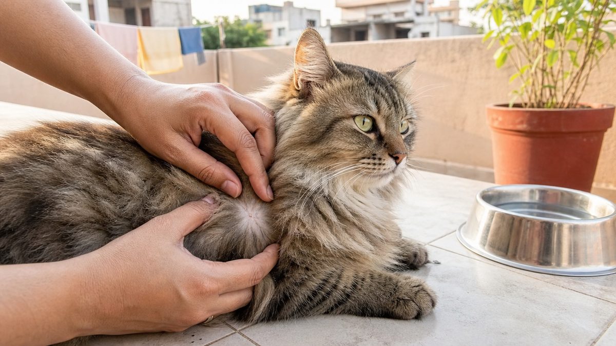
Quick answer
Ringworm in cats is a fungal infection of skin, hair and nails, not a worm, usually caused by Microsporum canis and passed to people as well as to other pets. It is self-limiting in many animals yet is always treated, because treatment shortens the contagious period. A clear Wood's lamp does not rule it out, culture and PCR confirm it, and a culture at a Chennai state veterinary hospital is priced at Rs 50.
This guide is general information for Indian pet owners and is not a diagnosis. Always consult a registered veterinarian near you before acting on anything you read here, and treat any sudden change in your pet's condition as urgent.
Signs of Dermatophytosis
The 10 signs to watch for are listed below. Any one of them is a reason to consult your nearby vet; none of them is a diagnosis.
- Circular patches of hair loss, often with scale at the edge
- Broken, stubbly hairs rather than a clean bald patch
- Grey scale deep in the coat, like cigarette ash
- Crusting on the face, muzzle, ear tips or paws
- Hair loss along the back, chest or forelegs
- Claws that turn rough, pitted, scaly or deformed
- A raised, weeping nodule on the muzzle or a foot
- A kitten or puppy losing hair in scattered patches
- A second pet, or a person at home, developing a ring
- No itch at all, which does not rule ringworm out
Breeds on this site that document this condition
Each breed page below carries the figure for this condition with its denominator and its source. The 9 breeds listed are the ones on this site that document it, not a ranking of risk.
India changes three things about ringworm. The species mix is not the Western one: Microsporum gypseum, a soil fungus, was 41 of 45 positive dogs in semi-arid Jaipur, while Microsporum canis was 29 of 36 isolates in Kerala cats and 60 percent of isolates around Kolkata. Healthy Indian pets carry it in numbers, 253 of 1,209 dogs and 109 of 292 cats near Kolkata. India is also where terbinafine-resistant Trichophyton indotineae emerged in people, and Indian canine strains of it have now been sequenced. A fungal culture at the Madras Veterinary College Teaching Hospital is priced at Rs 50.
Ringworm is a fungus, not a worm, and it is the commonest fungal skin disease cats get
Ringworm is a fungal infection of skin, hair and nails, and no worm is involved at any stage. The name describes the round, red, raised ring that marks the edge of the inflamed patch in people, and it is misleading twice over, because the infection is not caused by a worm and the affected areas are not always ring-shaped. The organisms are a specialised group of fungi called dermatophytes, which is why the medical name is dermatophytosis.
Three genera of fungi are collectively called dermatophytes, and the Government of Assam's own animal husbandry page names them as Epidermophyton, Microsporum and Trichophyton, noting that Epidermophyton occurs only in human beings while Microsporum and Trichophyton occur in cattle, sheep and goat, horse, pig, dog and cat. In companion animals the three that matter are *Microsporum canis*, *Microsporum gypseum* and *Trichophyton mentagrophytes*.
In cats, dermatophytosis is usually caused by *Microsporum canis*, is the most common fungal infection worldwide, and is one of the most important infectious skin diseases of the species. Severe disease sits mostly in kittens or immunosuppressed adults, poor hygiene is a predisposing factor, and the disease can become endemic in shelters or catteries.
Nobody can tell you how common it is, and the guideline says why
The true prevalence of ringworm is unknown, and the World Association for Veterinary Dermatology sets out exactly why in its clinical consensus guidelines. Determining true prevalence and breed predispositions is difficult because this is not a spontaneously occurring disease, it is not reportable and it is not a fatal disease, and infection varies in severity and resolves without treatment in many dogs and cats, all of which biases the breed and prevalence data.
So every percentage on this page, including the Indian ones, is a share of some clinic's caseload rather than a risk for a healthy animal, and the population is named on each. A 41.28 percent figure from Jaipur is 45 of 109 dogs already presented with alopecia, erythema and itching, and a 25 percent figure from Thrissur is 25 of 100 dogs already presented with hair loss, scaling and crusts. Neither is the chance your dog gets ringworm this year.
It is often self-limiting, and that is not a reason to skip treatment
Both halves of this are true at once and most pages carry only one. In most immunocompetent hosts dermatophytosis is a self-limiting skin disease within weeks to months, and the guideline still recommends treatment, with the stated goal of shortening the course of the disease to prevent spread to other animals and people. In many cats the disease shows hair loss and scaling only, and resolves by itself.
The page ranking first on Google India for this query puts the same point in one sentence, that although ringworm is self-limiting in many cats, treatment is always necessary to minimise the risk of spreading infection to humans, especially children, and to other pets. Waiting it out is a decision you are making on behalf of everyone else who lives in the house.
The one thing not to get wrong. Never put a combination cream containing a steroid on a suspected ringworm lesion, whether it is a human tube from the chemist or anything not dispensed for this animal. Topical corticosteroid use in tinea was strongly discouraged by all seventeen experts on the Indian dermatology task force that wrote the national consensus, and the earlier Indian expert consensus strongly discouraged it too. A steroid calms the redness, the lesion stops looking like ringworm, and the fungus keeps going. Take a spreading circular lesion to your nearby vet and ask for a culture rather than treating it blind. Find one through the Bonbeno vet directory.
What ringworm looks like on a cat and on a dog, and the things it is mistaken for
Ringworm looks like a circular patch of broken, stubbly hair with scale, and in cats it can look like almost nothing at all. Ringworm can be challenging to detect in cats because the lesions may be very mild or even undetectable, and a cigarette-ash scaling in the depths of the coat may be the only visible indicator. In cats the main sites are the skin of the head, chest, forelegs and along the ridge of the back.
The classic description is circular alopecia and desquamation, sometimes with an erythematous margin around a centrally healing patch, which is where the word ringworm comes from. Kittens are affected most commonly, and typical lesions are focal alopecia, scaling and crusting, most often around the ears and face or on the extremities, while lesions in dogs are classically alopecic, scaly patches with broken hairs.
Itch is not a reliable signal in either direction. Pruritus was reported in 63 percent of the 54 affected cats in a nine-year Italian referral series, regardless of breed, so an itchy cat is not thereby a flea-allergy cat, and a comfortable cat is not thereby clear. Claws can be involved on their own, turning rough and pitted with a scaly base and eventually deforming.
Widespread, crusty disease that will not settle is a signal to look for a second illness underneath it. Six Yorkshire terriers seen over three years with generalised, chronic *Microsporum canis* dermatophytosis all had a concurrent disease, four with leishmaniosis, one with leishmaniosis and ehrlichiosis and one with diabetes mellitus, and despite appropriate systemic and topical antifungal treatment only partial resolution was achieved, while the diabetic dog's infection resolved once the diabetes was stabilised. In cats, severe clinical signs are seen mostly in kittens or in immunosuppressed adults.
How ringworm is transmitted, and what carries it between animals
How ringworm is transmitted decides who else in the house needs looking at. The infective form is the arthrospore, formed by fragmentation of fungal hyphae into very small infective spores, and it moves by direct contact between an infected and an uninfected animal or by fomite transmission on grooming appliances, bedding, collars, ectoparasites and a contaminated environment, with concurrent microtrauma to the skin an important factor in whether an exposure becomes an infection. Contact with the fungus does not always result in an infection.
Age is the strongest signal you can read without a test
Young animals get it, and three separate series agree. Kittens were eight times more likely than adults to present with dermatophytosis at a Northwestern US shelter, with a 95 percent confidence interval of 4.8 to 13.5. In Thrissur the occurrence was highest in puppies under six months at 48 percent, against 36 percent between six months and two years and 16 percent from two to six years. In Jaipur, dogs between 6 and 18 months showed 46.67 percent against 31.11 percent above one and a half years and 22.22 percent over three years.
Animals under one year were also significantly more likely to be positive in a 150-animal Turkish sample, with p equal to 0.0097, and young age was a predisposing factor in the Italian referral series, where contagion in pure-bred kittens most likely occurred at the breeder's facility. A new kitten from a breeder or a litter off the street is the highest-probability starting point in an Indian household.
Four things that look like ringworm and are not
A ring-shaped lesion is a pattern, not a diagnosis, and four published alternatives sit behind it. The list below gives each one with its source.
- Figurate erythema in hairless cats. Eleven Eastern European sphynx-type cats, seven of them Donskoys, showed ring-shaped or serpentine red plaques with scaling trailing the expanding edge; dermatophytosis was ruled out by special stains or fungal cultures in eight of them, and no diet change or medical treatment achieved remission.
- Erythema annulare centrifugum in hairless dogs. A three-dog case series in two Peruvian Hairless and one Chinese Crested described ring lesions with clinical and histological features suggestive of the condition, resolving completely on oral or topical glucocorticoids, which is the opposite of what a fungus does.
- A non-dermatophyte fungus, or nothing at all. Of 100 Thrissur dogs presented with hair loss, scaling and crusts, only 25 grew a dermatophyte, while 60 grew *Aspergillus*, *Trichoderma*, *Sporothrix*, *Fusarium* or *Curvularia* and 15 grew nothing.
- A dermatophytic nodule rather than a flat ring. Subcutaneous dermatophytic infections are reported most commonly in Persian cats and Yorkshire terrier dogs, so a lump is not a reason to rule ringworm out.
The practical rule is that nothing on this list can be told apart by looking, which is the whole argument for the tests in the next three sections. Ringworm, flea allergy, mange and over-grooming all look similar to an owner and are treated completely differently.
What a Wood's lamp can and cannot tell you, with the numbers behind both claims
A Wood's lamp can find ringworm quickly and cheaply, and it cannot rule ringworm out. The fluorescence of infected hairs is apple green, it is strikingly distinct, and in active infections it involves the intrafollicular portion of the hair shaft, which can be examined grossly to confirm fluorescence and microscopically to confirm infection. It is the hair shaft that glows, not the scale and not the skin.
Lint, topical medicaments, seborrhoeic material and even soap residue will fluoresce under the lamp, but they lack the apple green or emerald green fluorescence of *Microsporum canis*. The original descriptions of the technique stressed holding the lamp close to the skin, 2 to 4 cm, to minimise false fluorescence and to concentrate on examining hair shafts. A lamp waved over a cat from arm's length is not the test.
The famous 'fewer than half of strains fluoresce' claim, traced to its four sources
The widely repeated line that under half of *Microsporum canis* isolates fluoresce comes from four laboratory studies, and the guideline went and found all four. Their range is 30 to 54 percent. The table below gives each one with its denominator, which no consumer page does.
| Study and population | Wood's lamp positive | What kind of number it is |
|---|---|---|
| Kaplan 1958, 2,183 dog and cat specimens sent to a diagnostic laboratory over 18 months, of which 445 were culture positive for *Microsporum canis* | 30 percent of infected hairs | A laboratory's reading of submitted hairs, not a clinician's examination of an animal |
| Wright, a 20-year retrospective laboratory report, n = 300 culture-positive *M. canis* specimens | 32 percent | Laboratory specimens again, with no record of technician training or lamp type |
| Sparkes 1993, a retrospective laboratory study spanning 1956 to 1991, n = 1,368 positive cultures | 54 percent of cat hair and 38 percent of dog hair | The same study put the lamp's positive predictive value at 90 percent and negative predictive value at 94 percent |
| A fourth laboratory study, 424 clinical samples with 77 of 99 culture positive for *M. canis* | 37 of 77, which is 48 percent | Laboratory samples, and none of the four studies recorded the examination procedure or type of lamp used |
| Pooled first examinations of animals with naturally occurring disease, 15 studies | 72 percent, which is 378 of 523 animals | Animals examined by clinicians rather than hairs read in a laboratory |
| Untreated cats, across the same pooled set | 91 to 100 percent | Fluorescence is far higher before treatment starts; in previously treated animals it fell to 39 to 53 percent |
| Cats with experimentally induced disease | 100 percent, which is 190 of 190 | Expected, because the investigators were searching for a known infection |
The guideline's own conclusion inverts the folklore. Contrary to what is believed, Wood's lamp examination is likely to be positive in most cases of *Microsporum canis* dermatophytosis, fluorescing hairs are most likely to be found in untreated infections, and false results are most commonly caused by inadequate equipment, lack of magnification, patient compliance, poor technique or lack of training.
What the lamp measures when somebody actually measures it
Three studies have put a sensitivity and a specificity on the lamp against culture, and they do not agree with each other. At a Northwestern US shelter the Wood's lamp had a sensitivity of 66.8 percent, 95 percent confidence interval 60.2 to 73.4, and a specificity of 74.8 percent, 95 percent confidence interval 64.2 to 85.1, compared with dermatophyte test medium culture. In a Czech shelter of 70 cats the sensitivity was 71 percent and the specificity 92 percent, with culture prevalence 64.29 percent against 48.57 percent showing fluorescence.
The third measurement is far lower and it was made on cats with no lesions. In 127 Thai cats screened for subclinical infection, the Wood's lamp sensitivity for *Microsporum canis* was 37.5 percent, 95 percent confidence interval 21.2 to 57.3, against a specificity of 96.1 percent, 95 percent confidence interval 90.4 to 98.5. Read those three together and the pattern is that the lamp finds lesional disease reasonably well and misses silent carriage badly.
One more finding from the shelter data is worth an Indian multi-cat household. In 78.8 percent of littermate or household groups where the Wood's lamp results were mixed, every cat in the group turned out to be culture positive. If one kitten in a litter glows, treat the finding as a statement about the litter.
What to ask for. A Wood's lamp examination is a point-of-care test that costs almost nothing and takes a minute, so there is no reason to skip it. Ask your vet to look at hair shafts up close in a dark room, and to pluck any apple-green hair for microscopy and culture rather than just recording a glow. A negative lamp is not a clear result, it is a reason to culture.
Fungal culture and PCR, and the test that still says yes after the fungus is dead
Fungal culture and PCR are the tests that confirm ringworm, and neither is a gold standard. No one diagnostic test was identified as a gold standard by the World Association for Veterinary Dermatology, which instead recommends a combination of complementary tests: the Wood's lamp and direct examination to document active hair infection, dermatophyte culture by the toothbrush technique to identify the species and monitor response, and biopsy with special fungal stains for nodular or atypical lesions.
The guideline is blunt that the usual framing is wrong. Fungal culture merely detects the presence or absence of fungal spores on the hair coat or hair sample, and false positives and false negatives both occur. The right questions are which tests confirm an active infection so an informed decision can be made, and which tests confirm the absence of one.
The toothbrush culture is so sensitive it finds animals that are not infected
This is the single most useful number on the whole diagnostic question and no owner page carries it. Among 5,644 cats screened at a shelter by toothbrush culture, 10.4 percent were culture positive, and when all the clinical data were examined only 1.67 percent actually had disease, while the other 8.8 percent were exposed animals mechanically carrying spores on the coat without a clinical infection.
Six of every seven positive cultures in that population were fomite carriage rather than disease. That is why a vet may want a Wood's lamp finding or hair microscopy alongside the culture before starting weeks of treatment, and it is why a single positive culture on a healthy in-contact cat is not, on its own, a diagnosis.
Collection technique matters more than the brand of medium. Individually wrapped soft-bristle toothbrushes are mycologically sterile, the technique is simple, atraumatic, economical and fast, and false negatives happen most often when lesions are not sampled thoroughly or when the laboratory does not know how to inoculate a toothbrush sample and randomly plucks hairs off the bristles. A sterile carpet square works just as well: across 78 cultures from 39 Persian cats in a cattery, the carpet and toothbrush techniques agreed 100 percent of the time.
How long a culture takes, which decides how long you wait
Cultures are faster than the three weeks owners are usually quoted, at least for *Microsporum canis*. Under consistent incubation conditions, all 202 diagnostic dermatophyte test medium plates from infected cats showed recognisable colony growth before 7 days, with a median of 4 days, and all 19 plates from fomite carrier cats grew before 12 days, with a median of 5 days. A positive culture can sometimes be confirmed within a couple of days, though slow-growing spores can take up to three weeks.
PCR detects dead fungus, and that changes when treatment stops
This is the fact that decides the end of treatment and the ranking pages do not carry it. PCR detection of dermatophyte DNA can be helpful, but a positive PCR does not necessarily indicate active infection, because dead fungal organisms from a successfully treated infection will still be detected on PCR, as will non-infected fomite carriers. A positive PCR three weeks into treatment can be a report on a fungus your treatment already killed.
The guideline is equally precise about the other direction, and this is the part that helps you stop. A negative PCR in a treated cat is compatible with cure, and a negative fungal culture from a cat with no lesions and a negative Wood's lamp, allowing for persistent glowing tips, is also compatible with cure. Those glowing tips are a known finding in cats under treatment or after cure, because as the hair grows the new intrafollicular portion is non-fluorescent while strong fluorescence remains on the distal shaft.
On raw accuracy from a single sample set, PCR wins. Across 48 feline samples compared head to head by dermoscopy, direct hair examination, three culture media and PCR, direct examination and dermoscopy gave unsatisfactory results, Mycosel and Sabouraud were suboptimal, dermatophyte test medium was the most reliable traditional method, and PCR was the top performer on sensitivity, specificity and accuracy. The VCA page notes the other side, that PCR can give false positives and may fail to identify the species, which matters for tracing the source and judging the risk to people.
Species identification is not a luxury in India. Knowing whether you are dealing with *Microsporum canis* off another cat, *Microsporum gypseum* out of the soil or a *Trichophyton* species changes where the infection came from and who else is exposed, and the Indian regional data in the section below shows those three are not interchangeable.
Asymptomatic carriers, and why treating only the animal with the lesion fails
Asymptomatic carriers are animals with no lesion that still grow the fungus on culture, and they are the reason a household outbreak comes back. Many adult cats are asymptomatic carriers, and infectious arthrospores can survive in the environment for about a year, transmitted by contact with sick cats or healthy carriers and also on dust particles, brushes, clothes and other fomites.
The page ranking first in India tells owners to try to separate infected animals from non-infected animals and treat only the infected ones, adding that in some situations it may be preferable to treat all pets. The carrier data is what decides which of those two situations you are in, and that data is not on that page.
In India the carriage numbers in apparently healthy pets are high. In the largest Indian study of its kind, 1,501 healthy companion animals belonging to individual owners in and around Kolkata were examined between 2011 and 2013, and 253 of 1,209 dogs, which is 20.93 percent, and 109 of 292 cats, which is 37.33 percent, were positive for dermatophytes on direct examination, with *Microsporum canis*, *Microsporum gypseum* and *Trichophyton mentagrophytes* predominating. Those animals were healthy and their owners had no idea.
Coat length changes the carriage rate by eight times
Long hair is the single strongest lifestyle risk factor that has been measured. Among 127 cats screened by fungal culture, subclinical dermatophytosis reached 34.92 percent of the 63 long-haired cats, 95 percent confidence interval 22.94 to 46.90, against 6.25 percent of the 64 short-haired cats, 95 percent confidence interval 2.15 to 12.28, giving long-haired cats 8.05 times the odds, 95 percent confidence interval 2.44 to 33.97. Within that sample, 17 of 47 Persian cats, 36.17 percent, were positive against 2 of 50 domestic short hairs, 4.0 percent.
A separate Bangkok survey of 59 cats and their 59 owners found long-haired cats carrying *Microsporum canis* significantly more often, p equal to 0.024, and a significant association between cats and their owners for the same organism, p equal to 0.010. A Persian or Balinese in a humid Indian flat is the profile those two studies describe.
Carriage is not confined to hot climates or to pedigree cats. Six of 113 dermatologically healthy cats in Talca, Chile, 5.3 percent, carried a dermatophyte on the mat technique, all of them *Microsporum canis*, and confinement status was the only epidemiological variable associated with carriage, with indoor cats at 10.73 times the risk of outdoor cats. Every isolate in that study was resistant to fluconazole.
One hair on one chin contaminated two rooms
The guideline records a case that explains why a household outbreak behaves the way it does. Two rooms of shelter cats were fungal culture positive, and examination found one lesional, Wood's lamp positive cat in each room, each with a single infected hair on its chin, while all the cats and the whole environment had colony counts too numerous to count; after those two cats were removed and the rooms decontaminated, cultures from all the remaining cats were negative.
The good news attached to the same passage is that spores lying about do not keep reinfecting a treated animal. In several studies, persistent exposure to spores in the environment did not result in reinfection in cats that had been cured of infection, and infection from the environment alone is rare.
What this means for an Indian multi-cat home. If one animal is diagnosed, ask your vet about screening every in-contact dog and cat by toothbrush culture rather than treating only the one with the patch. A cat that looks perfectly well can be culture positive, and in the pooled shelter data most positives were fomite carriers rather than infected animals, so what you do with each result is a conversation with the vet who has examined the animals, not a rule you can read off a web page.
Treatment in general terms, and why topical and oral therapy go together
Ringworm treatment is an oral antifungal and a topical one used together, for weeks, with testing to decide when to stop. Successful treatment requires concurrent use of systemic oral antifungals and topical disinfection of the hair coat. This page names drug classes and never a dose, because the dose is set by the veterinarian examining your animal, against its weight, its liver and everything else it is taking.
Twice weekly application of lime sulphur, enilconazole or a miconazole and chlorhexidine shampoo are the topical therapies currently recommended as effective for generalised dermatophytosis in cats and dogs. Accelerated hydrogen peroxide products, climbazole and terbinafine shampoos show promise but cannot be definitively recommended until more in vivo efficacy studies exist.
Twice weekly is not a rounding of once weekly. In a study of 85 shelter cats treated with twice weekly lime sulphur and a three-week course of oral terbinafine, the mean time to mycological cure was 22.7 days, and when the rinses were reduced to once weekly the colony counts rose and the cats did not achieve cure until twice weekly application was resumed.
Lime sulphur outperformed the alternatives in a randomised trial
The most recent controlled evidence is a randomised, semi-blinded non-inferiority trial in four US shelters. Seventy-six cats with *Microsporum canis* were treated with oral itraconazole and randomised to lime sulphur, miconazole with chlorhexidine, or hydrogen peroxide shampoo, and lime sulphur significantly outperformed both alternatives on cure by seven weeks, with a mean time to cure of 27 days against 37 for miconazole chlorhexidine and 36 for hydrogen peroxide.
Inferiority of the alternatives was suggestive but inconclusive, because the confidence intervals around the difference of means included the inferiority margin, and shelter staff rated all three products the same for unpleasantness and difficulty. Younger age significantly prolonged treatment, while intake type, co-housing and low body condition score did not.
The Indian feline study reached the same conclusion independently, which is a genuine corroboration rather than one paper. Across 82 Kerala cats split into five treatment groups, the response was notably swifter with lime sulphur dip when only topical therapy was considered, and mycological cure was most rapid when lime sulphur dip was combined with oral itraconazole.
The oral drugs, by class, and what the guideline says about safety
Systemic antifungal drugs have a wide margin of safety. For systemic therapy in cats, itraconazole is the drug of choice and terbinafine an alternative, with the recommended topical treatment being repeated body rinses with enilconazole or with miconazole, with or without chlorhexidine. Which one your animal gets, and for how long, is a decision for your nearby vet.
Two things do not work and are still sold as though they do. Lufenuron has in vitro efficacy against dermatophytes but does not prevent or alter the course of dermatophyte infections, does not enhance the efficacy of systemic or topical antifungal treatment, and has no place in the treatment of dermatophytosis. Antifungal vaccines do not protect against challenge exposure but may be a useful adjunct therapy, and a safe and efficient vaccine for cats is not available.
Coconut oil and the home remedies, checked rather than dismissed
Google India asks whether coconut oil cures ringworm, so this page went and looked rather than waving it away. A Europe PMC search on 27 September 2026 for coconut with any dermatophyte term returned four records, and adding a dog or cat term returned zero, with a Labrador control returning 110 on the same route in the same session to show the query was running.
Of those four records, the only recent one is a 2025 laboratory evaluation of coconut oil chitosan nanoparticles against *Microsporum canis*, and the other three are a 2025 onychomycosis hydrogel formulation paper, a 1992 study of Indian hair oils inhibiting dermatophyte growth and a 1975 paper on hair oils and unsaturated fatty acids in experimentally induced dermatophytosis. Activity in a dish is not a treatment for an animal, and none of the four is a clinical trial in a dog or a cat.
Clipping, which most pages recommend and the guideline does not
Clipping the coat is not the automatic first step. No study was identified that specifically addressed whether to clip, clipping was mentioned in only nine of the 57 reviewed treatment studies, and in three of those studies clipping the coat resulted in spread of the infection. Clipping is stressful, may require sedation, risks microtrauma to the skin and thermal injury from over-used clippers, and in multi-cat situations can increase spread if precautions are not taken.
Set against that, clipping a long coat does reduce the material shedding into the room, and the VCA page recommends clipping the hair shorter than 2 cm and disposing of it carefully, while noting that clipping at a veterinary clinic is not generally recommended because of the risk of spread to other pets. Both positions are defensible and your vet is weighing the coat in front of them.
Do not stop when the coat looks better. Treatment usually lasts a minimum of six weeks and sometimes longer, and if therapy stops too soon the disease recurs. Two consecutive negative fungal cultures are what indicates successful treatment. Ask your vet at the first visit what the stopping test will be and book it, so the end of treatment is a result rather than a guess.
Environmental decontamination is half the job, and it is where households give up
Environmental decontamination is ordinary cleaning done thoroughly, and the evidence says it works. Its primary purpose is to prevent fomite contamination and false positive fungal culture results, infection from the environment alone is rare, and infective material is easily removed, because if it can be washed, it can be decontaminated.
The strongest single piece of evidence on this comes from 70 real family homes rather than a laboratory. Over a ten-year period, 70 foster homes where *Microsporum canis* infected cats had lived were decontaminated by removing mechanical debris, cleaning with over-the-counter household detergents, rinsing, and disinfecting hard surfaces with household bleach at a 1 in 100 dilution or with accelerated hydrogen peroxide.
Thirty-eight of the 70 homes were completely decontaminated after a single cleaning once the cat was removed or cured, and of the rest, decontamination was achieved after one more cleaning in 28 homes, two more in two homes and three more in one. One home could not be decontaminated and the foster family was admittedly non-compliant, and across all 70 homes there was no evidence of disease transmission to other animals or people. The author's conclusion is that decontamination is not difficult and there is a perception in the lay literature that it is impossible.
The order of operations matters more than the disinfectant
Disinfection of a hard surface is three steps and the first two do most of the work. Mechanical removal of all debris by vacuuming or sweeping comes first, because disinfectants will not work in the presence of organic debris; washing with a detergent until the area is visibly clean comes second, because detergent lifts debris off the surface and must then be rinsed away since some detergents inactivate disinfectants; the disinfectant itself is the third step and kills residual spores. Those first two steps alone will decontaminate a surface in many cases, as has been shown in shelters.
| Surface | What was tested | Result |
|---|---|---|
| Laundry (cotton, terry cloth, denim) | Fabrics contaminated with infective spores and hairs, washed at 30 or 60 degrees C, with and without sodium hypochlorite, with and without machine drying | Washable textiles were decontaminated by mechanical washing at any water temperature, sodium hypochlorite was not helpful, and two washings on the longest cycle were effective without overloading the machine |
| The washing machine and dryer themselves | Surfaces sampled after the laundry study | Minimally contaminated, and easily cleared by spraying with accelerated hydrogen peroxide |
| Carpet | Carpet exposed to infective hairs and spores, cleaned several ways | Vacuuming alone did not decontaminate it but is still recommended to lift gross debris and infective hairs; washing twice with a carpet shampooer and detergent, or hot water extraction, did decontaminate it, and hot water extraction dried fastest without discolouration |
| Wood floors | No safe surface disinfectant exists for them | Daily removal of hair and dust with disposable dry mopping cloths, then washing twice weekly with a wood oil soap |
| Household bleach as a disinfectant | Sodium hypochlorite from 1 in 10 to 1 in 100 dilution, tested for antifungal efficacy and for shelf life | Consistently effective even with short contact times, but a 5.25 percent solution diluted 1 in 100 and not stored in a brown opaque container retained only 40 to 50 percent of its chlorine after 30 days, so it should be made up at least weekly |
The guideline lists real reasons not to reach for bleach at all, including its lack of detergency, which is a critical factor for disinfection, its potential to react with other chemicals and create toxic gases, damage to hard surfaces and floor finishes, discolouration of fibres, rapid loss of efficacy once diluted, and irritancy to both animals and people.
How often, and how much of the house
Twice weekly cleaning and disinfection is the recommendation, based on shelter studies and on studies of homes where cats were treated, covering mechanical removal of hair, washing and disinfection of the target areas. Restricting the animal to rooms that are easy to clean is the other half, and it is worth doing.
Confinement has a cost the guideline insists you weigh. It needs to be used with care and for the shortest time possible, because dermatophytosis is a curable disease while behaviour and socialisation problems can be lifelong if a young or newly adopted animal is not socialised properly. Socialisation should begin at three to four weeks for kittens and three to five weeks for puppies, and kittens do best when it happens by nine weeks of age and puppies by twelve to fourteen weeks. Items in the confinement area should be limited to things that can be washed daily, and toys should be plastic.
One owner action reduces the cleaning burden more than any product. Two studies showed that topical therapy with twice weekly shampooing using chlorhexidine and miconazole prevented contamination of the home, so treating the coat is also a decontamination measure and not only a treatment.
The Indian floor problem, stated honestly. In a home where people sit, eat and sleep at floor level and share bedding with animals, the contact surface is larger than in the houses these studies were run in, and no published study has measured that. What transfers is that the two steps doing most of the work, mechanical removal then detergent washing, cost nothing but effort, and that washable textiles decontaminate in an ordinary machine at any temperature over two long cycles. Daily damp mopping of the rooms the animal uses is the version of that advice that fits an Indian house, and the VCA page recommends exactly that, damp mopping or vacuuming daily.
The risk to the people in the house, with the numbers that actually exist
Ringworm passes from animals to people, the disease it causes in people is treatable, and the true transmission rate is unknown. Dermatophytosis is a known zoonosis and causes skin lesions which are treatable and curable, it is a common skin disease in people, and the true rate of transmission from animals to people has not been established. Serious complications of animal to human transmission are exceedingly rare.
This page does not diagnose or treat people, and nothing below is medical advice for a human being. If anyone in the house develops a skin lesion, especially a small patch of thickened, reddened skin with a raised scaly edge, that person should see their own doctor.
Who is most at risk, named by the guideline
Risk is concentrated at the ends of life and in people whose immunity is compromised. Pet-associated illness can occur in anyone, but people under 5 years of age, people 65 and over, pregnant women and people with immunocompromising conditions are at greater risk, and dermatophytosis appears on the guideline's own table of zoonotic diseases most frequently listed as a concern for high-risk clients. Children under 5 are the most at-risk human patient group for dermatophytosis specifically.
The immunocompromised category is broader than most owners assume and includes congenital immunodeficiency, transplant recipients, HIV infection, diabetes mellitus, chronic kidney failure, splenectomy, cancers, and treatment with immunosuppressive or chemotherapeutic drugs. A household with a person on chemotherapy or a transplant recipient is a household where the vet should be told.
The transmission numbers, all four of them
Only a handful of studies have ever put a denominator on animal to human transmission, and they disagree in an instructive way. The list below gives each one as the guideline reports it.
- 50 percent, from a letter to the editor. Of 92 people in 23 households in contact with infected cats, 46 developed skin lesions. This is the figure most often quoted and it comes from correspondence rather than a prospective study.
- 25 percent of pets, looked at from the human end. Where owners had medically confirmed *Microsporum canis* tinea corporis, dermatophytes were isolated from 98 of 384 of their pets, and of 78 owners with confirmed lesions, 38 had a culture-positive pet at home, 8 dogs and 30 cats.
- 14.6 percent of cats whose owners had no lesions. *Microsporum canis* was isolated from 28 of 192 cats living with owners who had no skin lesions at all, while it was not isolated from any dog whose owner was lesion-free.
- The negative result inside the same study. *Microsporum canis* was not isolated from the coats of 14 of 22 dogs and 26 of 56 cats belonging to owners with confirmed infection, which the guideline reads as showing that the pet may or may not be the source of a human infection.
No study was found that prospectively followed transmission from an infected pet to disease-free owners. The honest summary is that living with an infected cat raises your risk, that the household pet is not automatically the source when a person gets tinea, and that nobody has measured the rate properly.
What the disease looks like in a person, and why it is usually not serious
In people the predominant dermatophyte pathogen is *Trichophyton rubrum*, which is not animal-derived, and the most common clinical presentation is onychomycosis, meaning nail infection. Reports of *Microsporum canis* infection in immunocompromised people are limited to single case reports of tinea capitis, pseudomycetoma or mycetoma; across 21 such cases in two reviews, animal contact was confirmed in only 7, none of the patients died of the dermatophyte infection, and the most common complication was a prolonged treatment time.
In a review of 84 patients with severe dermatophytosis and acquired or innate immunodeficiency, occurrence was rare, the most common pathogen was again *Trichophyton rubrum*, and the most common underlying conditions were solid organ transplant in 28, CARD9 deficiency in 19 and HIV in 9. The picture is a nuisance infection with a long treatment, not a catastrophe.
The Indian family case, which is the one to read
India has a published household case that shows exactly how this runs in a home with children. Three family members in Delhi were diagnosed with tinea capitis caused by *Microsporum canis*; the index case was a five-year-old boy who contracted it through contact with a cat, and his younger brother and sister then acquired it by human to human transmission within the family, with molecular typing confirming the isolates were clonal.
Two details in that case matter for an Indian reader. The isolates were sensitive to itraconazole and terbinafine but resistant to griseofulvin, and all three patients did well on treatment chosen by their own doctors. The authors describe *Microsporum canis* tinea capitis as rarely reported in a non-endemic region like India, which means an Indian doctor may not think of the cat first. Telling your doctor that there is a cat at home with a skin lesion is genuinely useful information.
The guidance gap, measured
A 2026 scoping review looked for evidence-based guidance on the zoonotic side and found almost none. Of 554 human medical and 137 veterinary publications screened, only five of each met the inclusion criteria, none used an evidence-based guideline approach such as GRADE or AGREE, a significant proportion gave limited supporting evidence for their recommendations, and there were significant gaps and minimal synergies between the veterinary and human medical recommendations, with the human literature carrying limited information on zoonotic recommendations at all.
Dermatophytosis is also the zoonosis most frequently contracted by veterinary professionals in the UK from their patients, which is worth knowing when you hand an animal over at a clinic. In Gujarat, a survey of 120 dog owners across Ahmedabad, Anand and Vadodara found that awareness of the zoonotic aspects of canine scabies and dermatophytosis correlated significantly with extension contact, mass-media exposure and management orientation, while education, years of dog keeping and housing space showed no significant relationship. Knowing about it is not a function of schooling; it is a function of somebody telling you.
What Indian studies actually grow, and why it is not the same fungus in every state
Indian isolate studies grow different dermatophytes in different states, and the difference changes where the infection came from. In semi-arid Jaipur the dominant organism in dogs was the soil fungus *Microsporum gypseum*, in coastal Kerala it was *Trichophyton* species in dogs and *Microsporum canis* in cats, and around Kolkata it was *Microsporum canis*. The table below sets the published Indian series side by side with their denominators.
| Study, place and year | What was sampled | What grew |
|---|---|---|
| Singathia, Jaipur, Rajasthan, 2014 | 109 dogs presented to a teaching veterinary clinical complex with alopecia, erythema and itching between June 2009 and May 2013 | 45 positive, 41.28 percent. *Microsporum gypseum* in 41 dogs (91.11 percent of positives), *Trichophyton mentagrophytes* in 3 (6.67 percent) and *Microsporum canis* in one (2.22 percent) |
| Tarra, Thrissur, Kerala, 2022 | 100 dogs presented with hair loss, scaling, crusts and desquamation between January and September 2021 | 25 grew a dermatophyte. Of those 25, *Trichophyton* species 68 percent, *Microsporum* species 28 percent and *Epidermophyton floccosum* 4 percent; 60 grew non-dermatophyte fungi and 15 grew nothing |
| Sheetal, Pookode, Kerala, 2024 | 82 cats presenting with dermatological lesions, cultured on dermatophyte test medium | 36 afflicted with dermatophytes, of which 29 were *Microsporum canis* and 7 were *Microsporum gypseum*, confirmed by PCR-RFLP |
| Murmu, Kolkata, West Bengal, 2015 | 362 clinically suspected cases, comprising 123 dogs, 202 cats and 37 human beings | 285 positive: 158 cats (55.5 percent), 108 dogs (37.8 percent) and 19 humans (6.7 percent). *Microsporum canis* 60.0 percent, *M. gypseum* 22.5 percent, *Trichophyton mentagrophytes* 15.8 percent, *T. rubrum* 1.7 percent, with incidence highest in the rainy season from April to August |
| Debnath, Kolkata, West Bengal, 2016 | 1,501 clinically healthy owned animals, 1,209 dogs and 292 cats, examined 2011 to 2013 | 253 of the dogs (20.93 percent) and 109 of the cats (37.33 percent) positive on direct examination, predominantly *M. canis*, *M. gypseum* and *T. mentagrophytes* |
| Ranganathan, Madras, Tamil Nadu, 1997 | 211 dogs, both strictly house dogs and strays, plus 170 cattle | 106 strains, 89 from dogs. *Trichophyton mentagrophytes* var. *mentagrophytes* 57 of 106 and the *Microsporum gypseum* complex 42 of 106, with *M. gypseum* predominating in strays and cattle and *T. mentagrophytes* and *T. rubrum* in strictly house dogs |
| Begum, Uttarakhand, 2020 | 157 clinical samples from mixed animal species over three years, December 2016 to December 2019 | Prevalence ranged between 45 and 53.3 percent, with *Trichophyton mentagrophytes* the most abundant at 38 percent followed by *Microsporum canis* at 21.5 percent |
For scale against a country that has counted properly, Portugal tested 4,716 samples from dogs and cats with suspected dermatophytosis submitted by 355 veterinary centres between 2012 and 2023, and of the 568 positive cultures *Microsporum canis* was the most frequently isolated species at 63.9 percent, followed by *Trichophyton* species at 20.3 percent and *Nannizzia gypsea*, formerly *Microsporum gypseum*, at 8.1 percent, with cats positive more often than dogs at 17.4 percent against 9.1 percent. India has nothing on that scale, and the largest Indian series is 1,501 animals in one city.
The species split on the Indian government page does not match the Indian studies
The Government of Assam's animal husbandry page ranks on the India query and states that ringworm in dogs is caused mainly by *Microsporum canis* in 70 percent of cases, with *M. gypseum* at 20 percent and *Trichophyton mentagrophytes* at 10 percent, and that in cats 98 percent are caused by *M. canis*. The page gives no source, no sample size and no denominator for any of those four percentages.
For cats the direction is broadly right, since 29 of 36 Kerala isolates were *M. canis*. For dogs the published Indian series do not support it: Jaipur grew *M. gypseum* in 41 of 45 positive dogs and *M. canis* in exactly one, Thrissur grew *Trichophyton* species in 68 percent of its 25 isolates, and Madras grew *T. mentagrophytes* in 57 of 106 strains. An Indian owner reading that page would be told to look for the wrong organism.
Why the soil fungus matters practically
*Microsporum gypseum* is geophilic, meaning it lives in soil, and it is widespread in warm, humid, tropical and subtropical environments. A dog that grew *M. gypseum* did not necessarily catch it from another animal, so the household logic changes: the reservoir is the ground the dog digs in, and screening every other pet is less useful than it is for *M. canis*. Working and hunting activity increases exposure to dermatophyte spores for the same reason.
Indian dogs have grown the human fungus, which is the direction nobody writes about
Transmission in India has been documented running from people to animals, twice. In Thrissur, *Trichophyton rubrum*, an anthropophilic species that infects humans and cannot survive in soil, made up 35.3 percent of the *Trichophyton* isolates from dogs.
The Madras survey worked out the same thing from the owners' side. The owners of 11 dogs that yielded *T. mentagrophytes* var. *mentagrophytes* had tinea corporis or tinea pedis themselves, the owners of 4 of the 5 dogs that yielded *T. rubrum* were known *T. rubrum* patients, and the authors concluded that reversed transmission of dermatophytes from humans to animals may explain why these species predominated in strictly house dogs, with the pet dogs presumed to be the source of the owners' reinfection. A household ringworm problem in India can start with a person.
Two Indian studies disagree about the season, and both are right about their own city
Season is not a national fact. In semi-arid Jaipur, 23 of the 45 affected dogs, 51.11 percent, presented during the pre-monsoon and monsoon months from June to October, and the authors also recorded that infection was more severe and more prevalent in pets kept indoors in cooler surroundings using desert coolers and air conditioners, because higher humidity favours faster multiplication of fungal elements.
In humid coastal Thrissur the pattern reverses, with occurrence higher during the summer season than during the monsoon or winter. Around Kolkata the incidence was highest in the rainy season from April to August. If you want a seasonal rule for your own city, the honest answer is that three Indian studies give three different answers and the baseline humidity of the place decides. Our monsoon care guide for dogs in India covers coat drying, which is the shared control in all three.
Persian cats, and the Indian study that names them
Persians are the breed the Indian veterinary literature singles out. In an ICAR Indian Veterinary Research Institute study that sampled 548 cats and dogs, *Microsporum canis* was isolated from 52 of 74 cats, 70.27 percent, against 8 of 474 dogs, 1.68 percent, and Persian cats were found to be highly susceptible to *M. canis* infection. Terbinafine was the most effective antifungal against those isolates in vitro and fluconazole the least.
There is a genetic explanation under that. Whole-genome sequencing of case and control Persian cats identified a highly divergent disease-associated haplotype on chromosome F1 containing the S100 family of genes, with 13 non-synonymous variants in *S100A9* between cases and controls, and calprotectin expression was markedly upregulated in the feline epidermis during dermatophytosis. Persians show severe forms of the disease more commonly than other cat breeds, including other long-haired breeds.
Trichophyton indotineae, terbinafine resistance, and what the animal evidence does and does not show
*Trichophyton indotineae* is a terbinafine-resistant dermatophyte that was first identified in India, and Indian dogs have now been shown to carry it. This is a human medicine story at its centre and this page stays out of human treatment entirely, but three animal-side questions are legitimate and the published answers exist. What follows separates what is established from what is not.
*Trichophyton indotineae*, previously called *Trichophyton mentagrophytes* internal transcribed spacer genotype VIII, is a newly defined species frequently reported from India, it causes dermatophytosis in humans, and some strains are resistant to terbinafine. It was first identified in India and has since been reported across Asia, the Middle East, Europe and the United States.
On the human side, the Indian dermatology profession has described what changed. The past six to seven years saw an unprecedented increase in recurrent, chronic, recalcitrant and steroid-modified dermatophytosis of glabrous skin in India, with a notable decrease in clinical responsiveness to commonly used antifungals given in conventional doses and durations. Seventeen dermatologists reached consensus on 90 of 132 statements, and topical corticosteroid use, whether alone or in a combination product, was strongly discouraged by all of them.
The earlier Indian expert consensus said the same, that current treatment recommendations appeared to have lost their relevance in the clinical scenario, and it too strongly discouraged topical corticosteroid use in the practice of tinea management. An Indian prospective multicentre series has since described erythrodermic dermatophytosis, meaning fungal infection covering the whole skin, developing on a background of steroid misuse.
The cost of that mistake has been measured in rupees, on people rather than on pets. An Indian cost-of-illness analysis comparing steroid-naive with steroid-modified dermatophytosis found average total treatment costs of Rs 2,172.42 and Rs 3,770.61 respectively, an additional 40 percent on average for patients who had used topical steroids, driven by the need for more consultations, more investigations for the atypical presentation, and longer treatment with higher-grade antifungals. Both figures survive the conversion check, at roughly USD 25.75 and USD 44.69 at 84.38 to the dollar and USD 29.36 and USD 50.95 at 74.
That is a human figure and this page does not transfer it to animals, because no equivalent veterinary analysis exists in India. The mechanism it prices is the one a vet is warning about when they refuse to hand over a combination cream, which is a steroid making the infection atypical, harder to recognise and longer to clear.
Question one: has T. indotineae been found in Indian animals? Yes.
Indian dogs carry it and the isolates have been sequenced twice. Screening 442 samples at the Indian Veterinary Research Institute, 386 canine and 56 feline, recovered ten *Trichophyton* isolates, of which 5 were *T. mentagrophytes* genotype VIII, meaning *T. indotineae*, 2 were *T. interdigitale*, 2 were *T. simii* and one was *T. quinckeanum*, and the authors concluded that the genotype considered an endemic and emerging human pathogenic clone in India is also prevalent in animals.
A 2025 whole-genome study went further. Two *T. indotineae* strains isolated from canines in northern India were sequenced de novo, giving a 22 Mb genome with roughly 6,800 protein-coding genes in both, and across 16 Indian strains the single nucleotide polymorphism difference between any two ranged from 8 to 257, indicating limited variation within the species. Critically, the core-genome phylogeny revealed two major clusters with no cluster indicating a specific host association.
Question two: were the Indian canine strains resistant? The sequenced ones were not.
The title of the 2025 genome paper says it plainly and it is the detail that gets lost in the retelling. The canine strains sequenced were terbinafine-sensitive, and their squalene epoxidase coding sequences carried no amino acid substitutions at the key positions 393 and 397, while a high frequency of substitution at position 397 was found across the terbinafine-resistant strains in the comparison set. The study's own conclusion is that genome composition is similar in canine and human strains irrespective of the resistance phenotype, and that the pathogen and genotypes circulating among humans and animals need continuous monitoring to determine the exact role of animals in transmission and endemicity in India.
A separate Chandigarh and Bareilly study sequenced 13 *Trichophyton* isolates, 9 human and 4 canine, and found low single nucleotide polymorphism differences between human and dog isolates and between two cities more than 350 km apart, which the authors read as suggesting zoonotic transmission alongside fast spread over large distances; the resistant *T. indotineae* strains carried the SQLE F397L substitution while susceptible ones carried S395P or a wild-type sequence. Genotyping 24 isolates by amplified fragment length polymorphism found minimal genetic variability between animal and human origin isolates, which the authors again read as highlighting the possibility of zoonotic transmission, with reduced susceptibility to terbinafine and griseofulvin.
Where the evidence stops, stated plainly. Genetic similarity between human and canine isolates is consistent with transmission and does not establish its direction or its frequency. The authors of all three Indian genome studies use the words possible and possibility, not demonstrated. Nobody has published a prevalence figure for terbinafine-resistant *T. indotineae* in Indian dogs or cats, nobody has published a study of whether Indian owners apply human steroid and antifungal combination creams to their pets, and this page will not guess at either. What is established is that the organism is in Indian dogs, that Indian canine and human strains are near-identical genomically, and that the two sequenced canine strains were sensitive to terbinafine.
What has been documented in animals elsewhere
Outside India, three animal reports exist and they are all recent. A 2.5-year-old stray dog in Iran with hair loss, crusting and redness grew *T. indotineae* confirmed by internal transcribed spacer sequencing, with a multidrug-resistant phenotype against terbinafine, itraconazole and other antifungals, while luliconazole remained active in vitro, and sequencing showed Phe397Leu and Ala448Thr substitutions in the SQLE gene. In Egypt, the first isolation of *T. indotineae* in the country came from a survey of 140 animals with skin lesions, 90 dogs and 50 cats, in which 47 of 90 dog samples (52.22 percent) and 35 of 50 cat samples (70 percent) were culture positive and *Microsporum canis* was the most significant isolate.
A global scoping review of reports from 2009 to 2024 puts the broader pattern in place. Dermatophytes tend to infect younger animals with long hair, *Microsporum canis* is the most commonly reported species and is linked to cats, in vitro resistance to griseofulvin and fluconazole has been documented in multiple zoonotic dermatophyte species, and resistance to terbinafine and itraconazole is a possibility although the number of reports is scarce. The review also names the clinical trap, that healthcare providers may empirically treat with corticosteroids or antibacterial agents due to its atypical inflammatory appearance.
India also has a published canine case where standard therapy failed. An 8-year-old intact male Spitz in Jammu had persistent alopecia, scaling, erythema and pruritus despite itraconazole, a miconazole shampoo and a terbinafine dusting powder, and marked clinical improvement followed only after antifungal susceptibility testing directed a switch to posaconazole. One case is not a trend, and it is a reason for an Indian vet to consider susceptibility testing when a dog fails a properly given course rather than simply repeating it.
What the search found and what it did not
A Europe PMC search run for this page on 27 September 2026 found 11 records in title and abstract for dermatophyte terms with a dog or cat term and India, and 17 on author affiliation, the two sets overlapping but not identical, with a Labrador control returning 110 on the same route in the same session to prove the query syntax was working. Searching the species across the whole literature returned 243 records, of which 27 carried an animal term.
Two of the 11 title-and-abstract records were not about companion animals at all, and the affiliation search surfaced papers the title search missed, including the Indian *Microsporum canis* virulotyping study and the Kolkata zoonotic dermatophyte series. That is the whole published Indian animal-side literature on this question, and it is eight or nine papers.
What the tests cost in India, from a hospital that prints its prices
A fungal culture at the Madras Veterinary College Teaching Hospital in Chennai is priced at Rs 50, and small animal registration there is Rs 100, valid for seven days only. Those are published figures from a state veterinary university's own fee page, not estimates, and they are the cheapest honest benchmark an Indian owner has for this work-up.
The same hospital is equipped for the job. Its Centralized Clinical Laboratory has bacteriology, fungal, virology and cytology sections, and it lists Dermatology among the referral units of its Medicine department. The laboratory receives more than 30,000 samples a year on average from the hospital's units, the university farms and the research centres.
The pharmacy line changes the arithmetic further. The Pharmacy unit of the Department of Clinics runs on an average annual budget of Rs 25,00,000, and drugs and fluids are procured and dispensed free of cost for the treatment of animals brought to the hospital. A government veterinary college is therefore a materially different proposition from a private clinic for a six-week course of treatment.
Both rupee figures survive the conversion check, which matters because a converted dollar price masquerading as an Indian one is a common error on pet-cost pages. Rs 50 is about USD 0.59 at 84.38 to the dollar and about USD 0.68 at 74, and Rs 100 is about USD 1.19 and USD 1.35 on the same two rates. Neither lands on a round dollar figure, so this is a genuine Indian rate card rather than a list converted from elsewhere.
What this page cannot tell you
No source found for this page prints an Indian retail price for a lime sulphur dip, for oral itraconazole or for a PCR panel, and none is invented here. The Europe PMC audit and a corpus search across the Bonbeno guide and condition pages found no Indian rate card line for a dermatophyte PCR and no published Indian price for a veterinary antifungal course. Ring two practices in your city, whether that is Mumbai, Bengaluru, Hyderabad or Pune, ask for the price of a fungal culture, a Wood's lamp examination and a six-week course, and get the answers in writing before you start. A government veterinary college in your state is worth ringing as the second call, because the Chennai fee table above shows what that comparison can look like.
One structural point is worth carrying into that phone call. The most expensive part of ringworm treatment in a multi-animal Indian household is usually not the drug but the repeat cultures used to decide when to stop, and at Rs 50 a culture those are not what makes the bill. Skipping them to save money saves almost nothing and costs you the only objective signal that treatment has worked.
Living with it, and the weeks nobody warns an owner about
Living with ringworm means six weeks or more of twice-weekly topical treatment, daily cleaning of a few rooms, and a stopping decision made on a test rather than on the coat. Treatment usually lasts a minimum of six weeks and sometimes longer, and if therapy stops too soon the disease recurs. Infected pets remain contagious for about three weeks if aggressive treatment is used, and longer if only minimal measures are taken.
Timelines from the treatment studies give a more precise sense of the road. In one shelter study of 58 cats on oral itraconazole with twice weekly lime sulphur, the mean number of days to cure was 18 and every cat was cured by day 49. In a second study of 90 shelter cats across three topical products, the median time to cure on the original lime sulphur formulation was 30 days with a range of 10 to 69.
Where the whole environment is controlled, even a larger outbreak resolves. A shelter that combined stringent environmental decontamination, stratifying animals by lesion and culture status, oral itraconazole and twice weekly enilconazole dips cured 24 clinically affected cats and 22 lesion-free carrier cats, all culture negative between 30 and 56 days after starting treatment.
How long will my cat be contagious, and what shortens it
How long will my cat be contagious is the question owners ask first, and the answer is about three weeks under aggressive treatment. Topical therapy is what shortens the contagious window rather than the oral drug alone, because it disinfects the hair coat and minimises contamination of the environment, and in one shelter protocol using twice weekly lime sulphur the cage-mate kittens and juvenile cats housed in direct contact with infected animals, 32 of them, never developed lesions or became culture positive.
What actually goes wrong in households
The failure modes are predictable and none of them is exotic. The list below names each one with the evidence behind it.
- Dropping topical treatment to once a week. Colony counts rose and cats did not reach cure until twice weekly application was resumed.
- Stopping when the skin looks normal. Two consecutive negative fungal cultures are what indicates successful treatment, not the appearance of the coat.
- Treating only the animal with the lesion. Around Kolkata, 253 of 1,209 clinically healthy dogs and 109 of 292 clinically healthy cats were dermatophyte positive.
- Believing a clear Wood's lamp. Its sensitivity for subclinical infection in one 127-cat series was 37.5 percent.
- Trusting a positive PCR to mean the treatment failed. Dead organisms from a successfully treated infection are still detected on PCR.
- Skipping the cleaning. Thirty-eight of 70 exposed homes cleared on one cleaning, and only one of 70 could not be cleared, in a household where the family admitted non-compliance.
Handling, hygiene and the people at home
Wear gloves when handling an infected animal and wash your hands thoroughly afterwards, and wash your hands and sanitise the surfaces the animal touched after every bath or treatment. When an infected animal is in a home with other pets and family members, the guideline says family members, especially children, need to be advised about handling and the risk of infection.
The other side of that advice is that the animal still needs to be handled, examined and socialised, and that confinement should be as short as possible because behaviour problems from missed socialisation are permanent and dermatophytosis is not. An animal shut in a bathroom for eight weeks to protect the household has been traded for a different lifelong problem.
Prevention, and the honest limits of it
There is no vaccine worth relying on. Few efficacy studies on prophylactic or therapeutic anti-*Microsporum canis* vaccines for cats have been published, and a safe and efficient vaccine is not available. Antifungal vaccines do not protect against challenge exposure though they may be a useful adjunct to treatment.
What does reduce risk is the ordinary work. Drying a long coat properly after a monsoon walk or a bath, checking the skin under the coat weekly on a long-haired cat given 34.92 percent of 63 long-haired cats carried a dermatophyte subclinically, culturing a new kitten or puppy before it joins a household with a small child or an immunocompromised adult, and taking a circular lesion to a vet within days rather than trying a cream first. Our guides on grooming a cat at home in India and monsoon care for dogs in India cover the drying routine in detail.
One last thing about scale. Dermatophytosis is a curable disease, serious complications of animal to human transmission are exceedingly rare, and across 70 exposed family homes there was no evidence of disease transmission to other animals or people at all. It is a six-week nuisance handled properly and a six-month one handled badly, and it is almost never a disaster.
What Google India asks about ringworm in cats and dogs
What kills ringworm fast on cats?
Lime sulphur dip applied twice weekly, combined with an oral antifungal, is the fastest combination anyone has measured. In a randomised trial of 76 shelter cats on oral itraconazole, lime sulphur significantly outperformed miconazole chlorhexidine and hydrogen peroxide shampoos, with a mean time to cure of 27 days against 37 and 36. Your vet sets every dose.
Can humans get ringworm from cats?
Yes, humans can get ringworm from cats, and children under five are the most at-risk group. In one report, 46 of 92 people across 23 households in contact with infected cats developed skin lesions. Serious complications are exceedingly rare and the disease in people is treatable. See your own doctor about a human lesion, not your vet.
How do you know if your cat has got ringworm?
You confirm it with a test, because looking is not enough. A grey cigarette-ash scale deep in the coat may be the only visible sign, and lesions can be undetectable. A vet uses a Wood's lamp on hair shafts, direct microscopy of plucked hairs, a toothbrush fungal culture and sometimes PCR, and no single one of those is a gold standard.
Can cats get ringworm from litter?
Contaminated objects can carry infective spores, so a shared litter tray is a plausible fomite, but infection from the environment alone is rare. Arthrospores transmit on dust, brushes, clothes and bedding and survive about a year. Concurrent microtrauma to the skin matters more than exposure alone, and one well-documented human case involved car upholstery.
What kills ringworm fast on dogs?
The same combination as in cats, an oral antifungal plus twice weekly topical disinfection of the coat, because successful treatment requires both together. Lime sulphur, enilconazole or a miconazole and chlorhexidine shampoo are the recommended topicals for generalised disease in dogs and cats. No product on a shop shelf substitutes for a diagnosis and a prescription.
Can coconut oil cure ringworm?
No published veterinary trial supports coconut oil for ringworm. A Europe PMC search on 27 September 2026 for coconut with a dermatophyte term and a dog or cat term returned zero records, with a Labrador control returning 110 on the same route. One 2025 laboratory paper tested coconut oil chitosan nanoparticles against Microsporum canis in a dish, which is not a treatment.
Is ringworm in cats and dogs actually a worm?
No. Ringworm is caused by fungi called dermatophytes, and the name describes the round, raised ring seen in infected people rather than any organism. The name is misleading twice over, because no worm is involved and the affected areas are not always ring-shaped. The medical term is dermatophytosis.
Does a clear Wood's lamp mean my cat does not have ringworm?
No, a negative Wood's lamp does not clear a cat. Its measured sensitivity against culture was 66.8 percent in one shelter series, 71 percent in another and only 37.5 percent for subclinical infection in a 127-cat study. It is a fast, cheap first look at hair shafts, and a fungal culture is what settles the question.
Why does my vet want to test the other pets when only one has a lesion?
Because healthy-looking animals grow the fungus. Near Kolkata, 253 of 1,209 clinically healthy dogs and 109 of 292 clinically healthy cats were dermatophyte positive, and many adult cats are asymptomatic carriers. Treating only the animal with the visible patch is how a household outbreak keeps restarting after each course finishes.
Is the ringworm in Indian dogs the same fungus as in Indian cats?
Often not, and the species changes by region too. In Kerala cats, 29 of 36 isolates were Microsporum canis, while in semi-arid Jaipur dogs the soil fungus Microsporum gypseum accounted for 41 of 45 positives and M. canis for one. That matters because a soil organism did not come from another pet in the house.
Is the resistant ringworm in Indian news coming from pets?
Nobody has shown that. Trichophyton indotineae has been isolated and genome-sequenced from Indian dogs, and canine and human strains are near-identical, which the authors call possible zoonotic transmission rather than proof. The two canine strains sequenced in the 2025 study were terbinafine-sensitive, and no prevalence figure for resistant strains in Indian pets exists.
How long is my cat contagious, and when can treatment stop?
Infected pets remain contagious for about three weeks under aggressive treatment, and longer if only minimal measures are taken. Treatment usually runs a minimum of six weeks. Two consecutive negative fungal cultures indicate success, and a negative PCR in a treated cat is also compatible with cure, because the coat looking normal is not a result.